Abstract
Coronary artery aneurysms (CAAs) are rare cardiovascular abnormalities often associated with conditions like atherosclerosis, connective tissue disorders, or vasculitis. We report a case of a 39-year-old woman presenting with palpitations and dysphagia, ultimately diagnosed with a giant aneurysmal left circumflex coronary artery using a multimodal imaging approach. Initial echocardiography identified a cystic lesion within the left atrium, prompting further evaluation with cardiac magnetic resonance (CMR), computed tomography (CT), and coronary catheterization. These modalities confirmed a large LCX aneurysm measuring 6.3 × 7.9 cm with left main coronary artery dilation. While surgical intervention was recommended, the patient opted for medical management. This case underscores the critical role of advanced imaging in diagnosing and managing complex cardiac conditions.
Introduction
Coronary artery aneurysms (CAAs) are serious cardiovascular conditions that need to be diagnosed and treated cautiously with giant CAAs being even rarer which is incidentally found in patients undergoing coronary angiography with a prevalence of less than 0.8% [ ]. Giant CAAs, defined as aneurysms >2 cm in diameter, are even rarer and often incidental findings. Although atherosclerosis, vasculitis, and connective tissue disorders are common etiologies, their pathogenesis remains poorly understood. Accurate diagnosis is essential, as these aneurysms carry a significant risk of rupture, thrombosis, or myocardial ischemia. Multimodal imaging plays a pivotal role in identifying and characterizing these lesions, including echocardiography, cardiac magnetic resonance (CMR), computed tomography (CT), and coronary angiography. This report highlights the case of a young woman presenting with nonspecific symptoms, emphasizing the value of advanced imaging in diagnosing a giant LCX aneurysm.
Case presentation
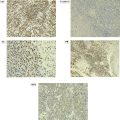
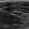
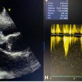
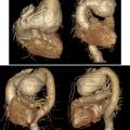
A 39-year-old woman presented with a 5-year history of intermittent palpitations, recently accompanied by dysphagia to solids. Physical examination and routine laboratory tests were unremarkable. An initial electrocardiogram (ECG) revealed sinus rhythm with a right bundle branch block, while transthoracic echocardiography (TTE) identified a cystic lesion in the left atrium measuring 6.5 × 7.3 cm, with blood flow detected by color Doppler ( Fig. 1 ). Further evaluation with cardiac magnetic resonance (CMR) demonstrated significant dilation of the left main coronary artery (8 mm in diameter) and a giant aneurysm of the left circumflex artery (6.3 × 7.9 cm), which filled during the perfusion study ( Fig. 2 ). Computed tomography (CT) corroborated these findings, revealing an LCX aneurysm measuring 6 × 7.5 cm and associated left main dilation ( Fig. 3 ). Coronary catheterization confirmed the LCX aneurysm and its connection to the left main coronary artery ( Fig. 4 ). A multidisciplinary team recommended coronary artery bypass surgery due to the aneurysm’s size and risk of complications. However, the patient declined surgical intervention and was managed conservatively with aspirin (81 mg daily), atorvastatin (40 mg daily), and bisoprolol (5 mg daily). She remains under regular follow-up, with no reported adverse events or progression of symptoms after 6 months.

Related posts:
 Breast cancer with medullary features shows a fast and plateau enhancement pattern on magnetic resonance images: A case report
 Ovarian metastasis from lobular breast carcinoma: A case report with review of literature
 Invasive lobular carcinoma with metastasis to the pectoralis muscle
 Early onset development of hypertrophic cardiomyopathy in less than 1 year in a patient with familial Friedrich’s ataxia: Case report
 Giant ascending aortic aneurysm: A rare case report
 A rare and life-threatening case of spontaneous hemopneumothorax presenting with severe hemodynamic instability
Stay updated, free articles. Join our Telegram channel

Full access? Get Clinical Tree


