Abstract
Acute scrotal pain includes urgent conditions in urology, such as testicular torsion, testicular rupture, epididymo-orchitis, and abscess. However, varicocele (pampiniform plexus) thrombosis is considered to be a rare cause of such pain. We herein report a case of a 27-year-old male patient with a history of epididymo-orchitis, who complained of painful scrotal swelling. Ultrasonography showed left-side pampiniform plexus thrombosis. This case highlights a rare condition, which should be included in the differential diagnosis of acute scrotal pain, indicating the need for further studies to elucidate its pathophysiology and provide proper treatment for such cases.
Introduction
Acute scrotal pain is a urological emergency; on the other hand, pampiniform plexus thrombosis is considered an extremely rare cause of acute scrotal pain. There are several possible predisposing factors for thrombosis, such as vascular endothelial injuries, sluggish venous flow, and hypercoagulability; however, infection has not been reported as the main factor. A few cases of thrombosed varicocele have been reported in patients with epididymo-orchitis. Alshubaili et al. [ ] published the first case in 2020. A second case was reported in 2021 by Hamdouni et al. [ ].
To our knowledge, this is the third case; however, it was on the left side , which is the more common side for varicocele rather than on the right side, as in the previous 2 cases.
Case report
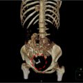
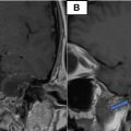
A 27-year-old single male patient, presented to the emergency department with a complaint of painful swelling in the left hemiscrotum for a few hours; the patient did not report any other symptoms. The patient was diagnosed with left epididymo-orchitis ( Fig. 1 ) and was started on oral antibiotics and analgesics for approximately 2 weeks. The patient had no significant medical or surgical history. Physical examination revealed swelling and tenderness in the left scrotum with no overlying skin changes. The laboratory results were unremarkable. The complete blood count was normal (erythrocytes: 5.28 × 10 12 /L, white blood cells: 8.1 × 10 9 / L [neutrophils 89%, lymphocytes 8%, monocytes 5%, eosinophils and basophils, 0%]). Scrotal ultrasonography was performed to rule out a strangulated inguinal hernia or abscess formation. Ultrasound revealed slight heterogeneity of the left testis of a normal size with an enlarged, heterogeneous left epididymis. In addition, dilated pampiniform plexuses with intraluminal echogenic content were observed, with no Doppler flow or pulse waves ( Fig. 2 ). The diagnosis based on imaging was thrombosed varicocele, and the patient was urgently referred to the urology team for further evaluation and treated surgically (varicocelectomy).


Related posts:
 Breast cancer with medullary features shows a fast and plateau enhancement pattern on magnetic resonance images: A case report
 A rare and life-threatening case of spontaneous hemopneumothorax presenting with severe hemodynamic instability
 Avascular necrosis of lateral femoral condyle in a middle-aged woman from central India
 A very rare case of ileocolic and appendiceal intussusception with acute appendicitis
 An incidental finding of an ectopic intrauterine device (IUD) in the urinary bladder wall in a female presented with missed abortion
 Petrous apex epidermoid cyst: A rare case
Stay updated, free articles. Join our Telegram channel

Full access? Get Clinical Tree


