KEY FACTS
Terminology
- •
Collagenous degeneration of rotator cuff ± biceps tendons with proteoglycan deposition
Imaging
- •
Supraspinatus > infraspinatus > biceps > subscapularis
- •
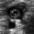
Diffusely thickened tendon with diffuse hypoechogenicity and indistinct fibrillar pattern
- ○
Graded as mild, moderate, or severe
- ○
- •
Cortical irregularity of tendon insertional area
- •
Biceps tendinosis usually accompanies rotator cuff tendinosis
- ○
Proximal end of bicipital groove is most common site of biceps tendinosis
- ○
- •
Subclinical tendinosis of similar severity often present on opposite side
- •
Ultrasound is better than MR for assessing tendinosis as tendon detail, such as fibrillar pattern, is better appreciated
- •
Calcific tendinosis: Focal echogenic mass within rotator cuff tendon with acoustic shadowing
Scanning Tips
- •
Heel-toe ultrasound probe to remove anisotropy artifact and avoid mistaking for tendinosis
- •
Tendinosis is associated with tendon tears and should also be assessed
- •
Tears are more linear, hypoechoic, and sharper in outline than tendinosis; may or may not be associated with volume loss of tendon contour
- •
Suggested protocol
- ○
Supraspinatus: Crass (arm extended and internally rotated) or modified Crass position (hand in back pocket)
- ○
Infraspinatus and teres minor tendons: Arm abducted and internally rotated (hand on opposite shoulder)
- ○
Subscapularis and long head of biceps tendons: External rotation of arm with elbow flexed
- ○
Confirm all findings in 2 planes
- ○


