Salivary Glands


Salivary Glands


Despite the realization of the diverse natural history and variation of radiation technique by specific site of origin, the rarity of salivary cancers led many investigators to report treatment outcomes in aggregate. Therefore, this chapter begins by summarizing the general background outcome data before addressing individual subsites.


Background Data


Table 13.1 Histologic Distribution of Salivary Neoplasms by Site


Site


Benign


Mucoepidermoid


Adenoid Cystic


Adenocarcinoma


MMT


Acinic


Epidermoid


Anaplastic


Parotid


1,342


272


54


62


107


75


45


8


Submandibular


106


37


45


9


24


2


8


Palate


60


37


67


41


18


1


4


Lip/cheek


13


23


12


20


2


3


Antrum


13


31


23


3


1


1


Tongue


2


14


30


12


2


3


Nasal cavity


4


12


17


23


1


3


Gingiva


13


10


6


3


1


1


Floor of mouth


1


6


7


8


Larynx


3


3


7


8a


Tonsil


4


3


3


1


2


Ethmoid


1


1


6


1


Nasopharynx


2


1


5


1


Pharyngeal wall


1


2


a All neuroendocrine carcinomas.


MMT, malignant mixed tumor.


From Spiro RH. Salivary neoplasms: overview of a 35-year experience with 2,807 patients. Head Neck Surg 1986;8:177-184, with permission.


Table 13.2 Local Failure of Adenoid Cystic Carcinoma Treated with Surgery and Postoperative Irradiation Stratified by Positive Margins and Named Nerve Involvement


Site


No. of Failures/No. of Patients (%)


No. of Failures/No. of Patients with Positive Margins (%)


No. of Failures/No. of Patients with Named Nerve Involvement (%)


Minor salivary gland


16/122 (13)


10/54 (19)


5/31 (16)


Submandibular/sublingual gland


1/41 (2)


1/11 (9)


1/14 (7)


Parotid gland


4/30 (13)


3/15 (20)


4/10 (40)


All patients


21/193 (11)


14/80 (18)


10/55 (18)


Data from the M.D. Anderson Cancer Center.


Modified from Garden AS, Weber RS, Morrison WH, et al. The influence of positive margins and nerve invasion in adenoid cystic carcinoma of the head and neck treated with surgery and radiation. Int J Radiat Oncol Biol Phys 1995;32:619.


Table 13.3 Five-Year Actuarial Local-Regional Control of Adenoid Cystic Carcinoma Treated with Neutron Therapy


Site


No. of Patients


Local-Regional Control (%)


Paranasal sinus


32


43


Oral cavity


26


68


Oropharynx


19


75


Nasopharynx


15


21


Submandibular-sublingual gland


15


59


Parotid


27


67


Modified from Douglas JG, Laramore GE, Austin-Seymour M, et al. Treatment of locally advanced adenoid cystic carcinoma of the head and neck with neutron radiotherapy. Int J Radiat Oncol Biol Phys 2000;46:551.


PAROTID


Treatment Strategy

Surgery is the preferred treatment for operable cases. Postoperative radiotherapy is indicated in the following clinical settings: high-grade tumors (mucoepidermoid carcinomas, malignant mixed tumors, adenocarcinomas, and squamous cell carcinomas); close or positive surgical margins (including incompletely resected recurrent pleomorphic adenoma); tumor adherence to or invasion of the facial nerve, or presence of histologic evidence of perineural spread; bone and/or connective tissue involvement; lymph node metastases (particularly with extracapsular extension); or after resection of recurrent disease even with negative margins.


Postoperative Radiotherapy


Target Volume


Initial Target Volume

The volume for benign and low-grade tumors without lymph node involvement is the parotid bed only. For high-grade tumors and tumors with lymph node involvement, the volume encompasses the parotid bed and ipsilateral neck nodes. For macroscopic invasion of the facial nerve, more generous coverage of facial canal to geniculate ganglion is desired.


The boost volume encompasses the tumor bed and the involved nodal bed.


Setup and Field Arrangement for Conventional Technique

Target volume 5 cm or less deep (superficial lobe and deep lobe in thin patients) (see Case Studies 13-1 and 13-2).

An intraoral stent containing cerrobend is used to shield the posterior oral tongue (see
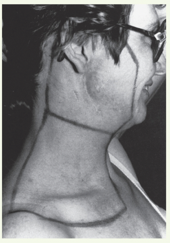
 Chapter 3).

Marking of the surgical scar and lateral canthus of the ipsilateral orbit facilitates portal design.

The patient is immobilized in an open-neck position with a thermoplastic mask. Flattening of the ipsilateral ear against the mastoid process minimizes dose heterogeneity resulting from electron perturbation. For the same reason, the external auditory canal is filled with Domeboro fluid prior to each electron treatment.


Bolus is used to cover the superior aspect of the portal when it extends above the zygomatic arch to minimize the dose to the temporal lobe of the brain.

A lateral appositional field is used to cover the parotid bed and upper neck nodes. Radiation is delivered with a combination of electrons (12 to 20 MeV depending on the depth) and photons (6 MV) usually in the ratio of 4:1.

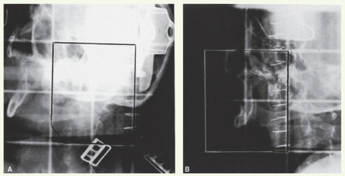
Portal borders are as follows:


  • Superior: zygomatic arch or higher as indicated by tumor extent or surgical scar.


  • Anterior: anterior edge of the masseter muscle.


  • Inferior: thyroid notch.


  • Posterior: just behind the mastoid process.

A 1.5- to 2-cm bevelled bolus or a computer-generated custom bolus is placed at the superior part of the portal (above the line connecting the orbital floor and the mastoid process) to reduce the dose to the temporal lobe. A lateral appositional electron field is used to treat the mid and lower neck nodes when indicated (for borders of neck field, see “General Principles”). Field reduction takes place after 50 to 54 Gy in 25 to 27 fractions to deliver the boost dose when indicated. If the anterior edge of the portal is close to the eye, skin collimation is applied and the beam maybe angled 5 to 10 degrees posteriorly to minimize the dose to the orbital content.

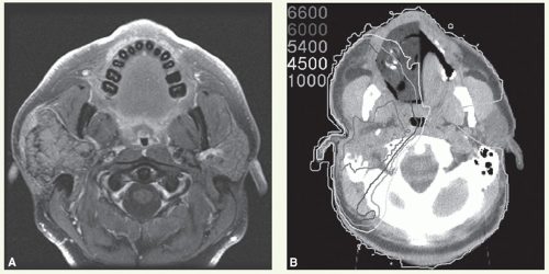
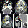
For more deep-seated tumors or when the facial canal is part of the target volume, a wedge-pair technique (see Case Studies 13-3 and 13-4) or intensity-modulated radiation therapy (IMRT) (see Case Studies 13-5, 13-6 and 13-7) with photon beams is often preferable.

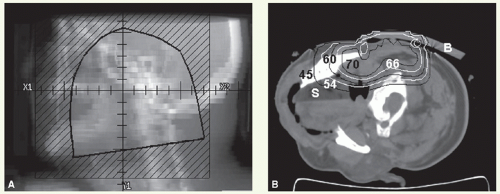
With these techniques, the patient is immobilized in a supine position with the head hyperextended with thermoplastic mask. The axial plane of the fields is chosen so that the posterolateral portal does not exit through the contralateral eye. A relatively simple wedge-pair technique uses anterolateral and posterolateral oblique photon fields (the anterolateral oblique field is on the spinal cord and the posterolateral oblique field is off the cord). The simulation focuses on marking of the surgical scar and both inferior orbital rims and selection of provisional isocenter. The provisional isocenter is generally placed at the center of the square defined by the zygomatic arch, anterior edge of the masseter, thyroid notch, and mastoid, and halfway between the skin and the oropharyngeal wall.

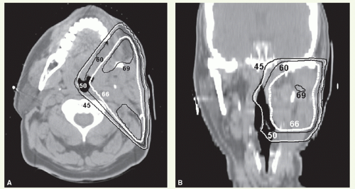
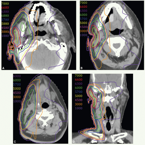
Thin-slice computed tomography (CT) scan is obtained in the treatment position for outlining the target volume and planning the portal sizes, hinge angle, and thickness of wedges using treatment planning system. No off-spinal cord reduction is required with a wedged-pair technique because the posterolateral field is off the cord from the beginning. Field reduction for boost dose, when indicated, occurs after 50 to 54 Gy.


Intensity-Modulated Radiation Therapy

With IMRT, the patient is immobilized with an extended head and shoulder thermoplastic mask in a supine position. Thin-slice CT scan is obtained for delineation of target volumes (see Case Studies 13-6 and 13-7).


Virtual Gross Target Volume

There is no actual gross target volume (GTV) after complete surgical tumor resection. However, it can be useful to formulate a virtual GTV (vGTV) to facilitate target volume definition. The vGTV is a best approximation of the
tissues having high likelihood of harboring microscopic tumor reconstructed based on findings of preoperative clinical examination, imaging studies, and surgical-pathologic assessment.


Jun 1, 2016 | Posted by in HEAD & NECK IMAGING | Comments Off on Salivary Glands

Full access? Get Clinical Tree

Get Clinical Tree app for offline access