Cases
19Isolated Fracture of the Anterior or Posterior Arch of Atlas
20Odontoid Fractures Types 1 and 3
21Unilateral Facet Dislocation
22Isolated Fracture of the Lamina
23Isolated Transverse Process Fracture
24Isolated Spinous Process Fractures
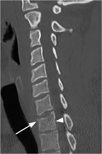
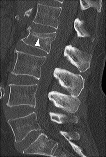
25Vertebral Body Microfractures / Bone Marrow Edema
Subsection 3BTypically Unstable
Subsection 3CSoft Tissue Injuries
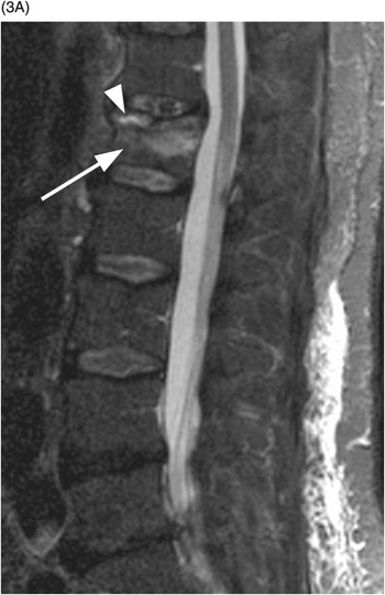
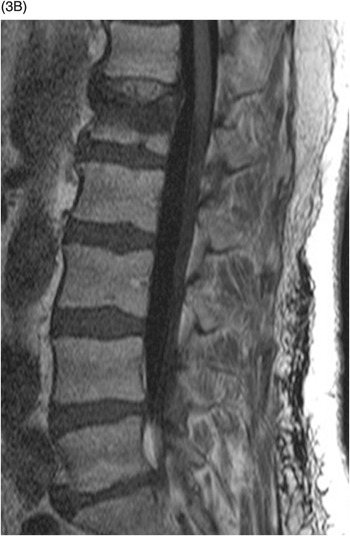
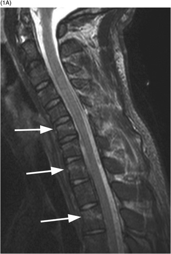
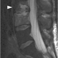
Figure 16.3 Midsagittal STIR MR image (A) reveals L1 superior endplate depression and underlying bone marrow edema consistent with a recent fracture (arrow). Note a small amount of fluid signal (arrowhead). The edematous bone marrow is hypointense on corresponding T1-weighted MR image (B). No retropulsion into the spinal canal.
Imaging Findings
Imaging reveals reduction of the anterior height and buckling of the superior-anterior part of the vertebral body due to impaction and cortical disruption of the anterior part of the superior endplate. There is often a horizontal sclerotic band in the upper vertebral body, reflecting trabecular impaction. Inferior endplate is affected less frequently. Posterior cortex is normal, as only the anterior column is involved. Most of the wedge fractures are symmetric, but in 8–14% of cases there is left–right asymmetry (lateral wedge fracture). There is focal kyphosis at the level of injury. MR imaging additionally shows marrow edema of the superior part of the vertebral body (T2-weighted sequences with fat suppression – T2 FS or STIR), possibly with fluid signal within the fracture. There is no anterior or posterior translation of the vertebral bodies. Underlying metastases or other neoplasm and osteomyelitis may be DWI hyperintense and without fluid signal on STIR images.
Differential Diagnosis
Unstable Wedge Fracture
Trauma to the anterior column and the posterior column, including posterior ligamentous complex – signs of posterior column disruption on CT and MRI.
Burst Fracture
Compression of whole vertebral body with posterior cortex involvement, often with sagittal fracture lines and fragmentation with posterior fragment dislocation – consider if loss of vertebral body height is more than 40%.
Chronic Wedge Fracture
An old fracture will have a certain degree of remodeling and smoothing of the cortical edges, without a clearly visible fracture line. Recently fractured cortex of the vertebral body is sharply marginated and often angulated. No bone marrow edema on MRI.
Scheuermann’s Disease
Most frequent thoracic (type I), followed by thoracolumbar (type II) – anterior wedging of at least three contiguous vertebral bodies; endplate irregularities are always present; no anterior vertebral body buckling.
Clinical Findings, Implications, and Treatment
The main symptom is pain; in osteoporotic fractures the onset may be insidious and the pain may be mild. As this type of fracture does not involve retropulsion of bone fragments into the spinal canal, neurologic symptoms are rarely present.
Stable wedge fractures make up for 50% of all traumatic thoracolumbar fractures, most commonly occurring at levels T12–L2. In the setting of osteoporosis insufficiency, compression fractures can occur at any level, most frequently in midthoracic spine. Nonoperative treatment is the standard: it includes pain medication and brief rest. Early mobilisation is recommended, using hyperextension braces for 6–12 weeks. Vertebroplasty or kyphoplasty is considered, especially in patients with long-standing pain or severe kyphotic deformity.
Additional Information
The presence of injury to the anterior longitudinal ligament, superior endplate and disc, or a high level of bone edema appear to be the critical factors that determine progression of kyphotic deformity following conservatively treated stable thoracolumbar compression fractures. The intraosseus fluid sign on CT can be seen in acute (as well as chronic) fractures and seems to predict greater height loss and kyphotic angulation.
References
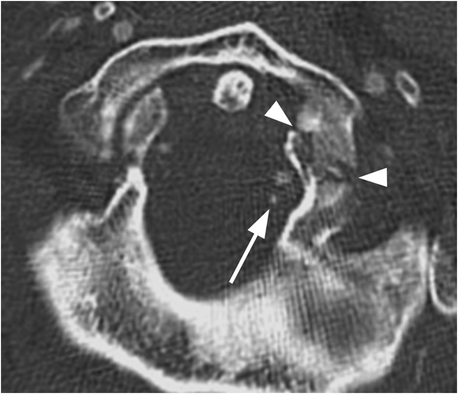
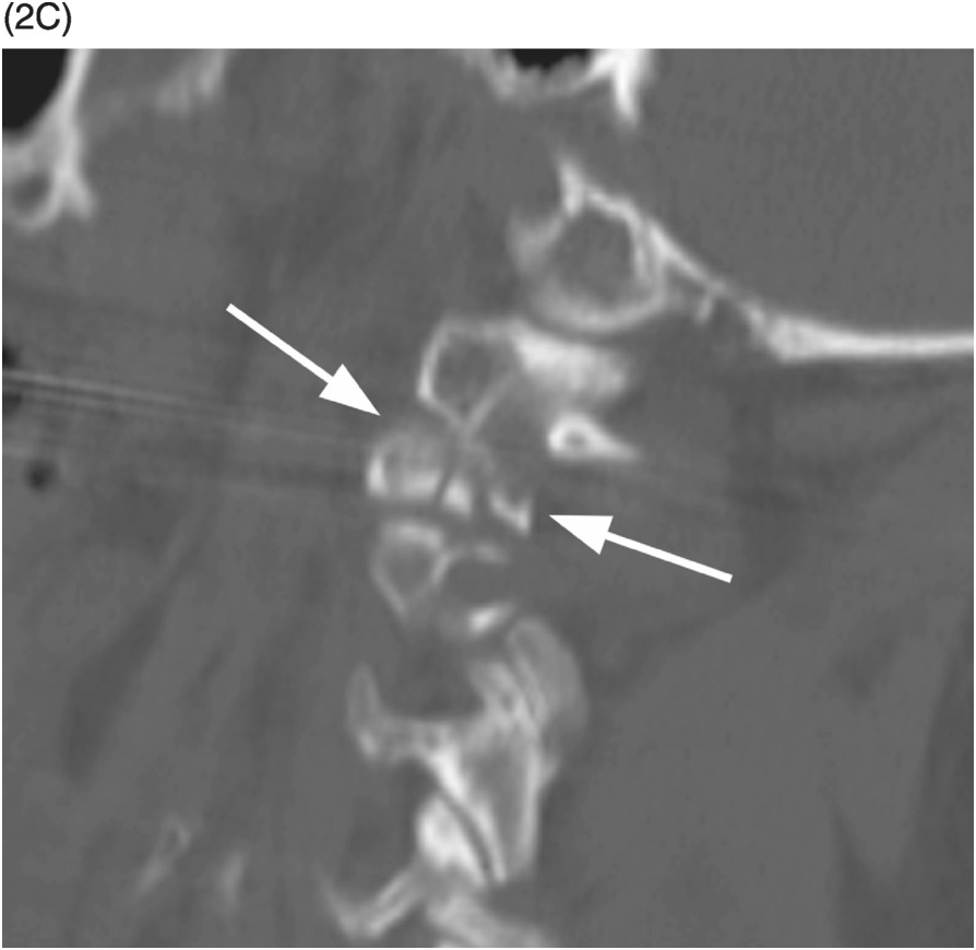
Figure 17.3 Axial CT shows an antero-posteriorly directed fracture of the posterior inferior occiput extending into the foramen magnum. There is a subtle fracture line of the right occiput (arrow) with vertical extension more inferiorly into the right occipital condyle. This is Anderson Type II occipital condyle fracture.
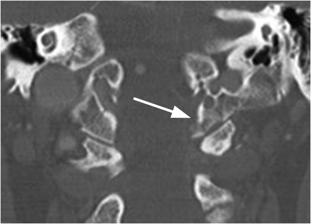
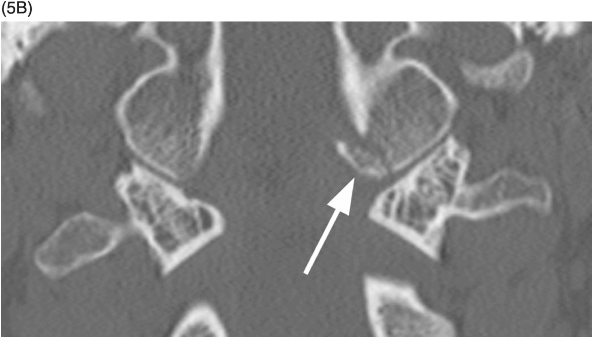
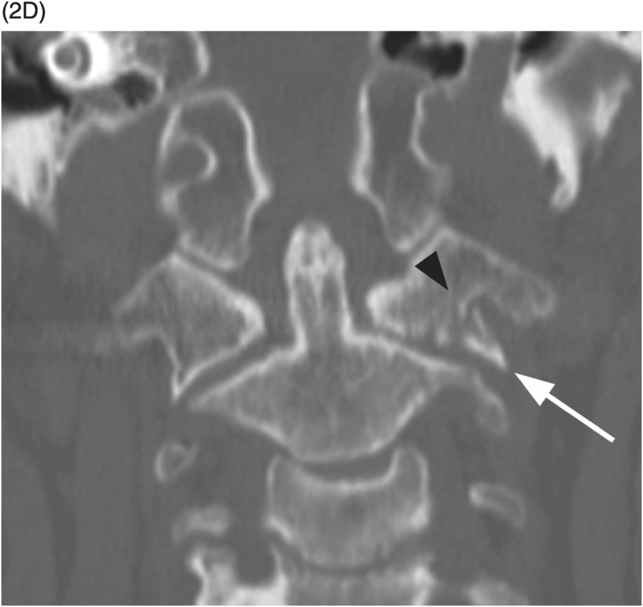
Figure 17.4 Coronal CT image shows an avulsion fracture of the inferior medial aspect of the right occipital condyle (arrow) at the insertion site of the right alar ligament. This is an Anderson Type III fracture, the least stable of the condyle fractures, but is generally treated conservatively as well. Note that there is no widending of the atlanto-occipital joint.
Imaging Findings
Occipital condyle fractures need to be evaluated with multiplanar CT, as they may at times be clearly seen in a single plane only. These fractures are classified into three types by the Anderson and Montesano scheme: Type I – comminuted and nondisplaced secondary to axial loading; Type II – extend into the condyle from a linear fracture in the remainder of the skull base (generally involve the base of the occipital condyle without complete separation of the condyle from the skull); Type III – avulsion fractures of the occipital condyle. Tuli et al. proposed a classification scheme consisting of type 1 (nondisplaced), type 2a (displaced stable), and type 2b (displaced unstable) fractures. This group suggested that more than 8 degrees of rotation or 1 mm of translation of the occiput relative to C1, direct evidence of alar ligament avulsion, or MRI evidence of atlanto-axial disruption is consistent with instability. Anderson classification is more widely accepted.
The role of MRI for direct inspection of the alar ligaments and tectorial membrane is questionable. More important is careful inspection of CT images for additional injuries of the skull, atlas, and axis because of the significant forces involved and anatomic proximity of critical structures.
Differential Diagnosis
Clinical Findings, Implications, and Treatment
Because of the associated high-energy mechanism of injury, patients with occipital condyle fractures frequently are obtunded at presentation; otherwise, pain is the predominant complaint. Lower cranial nerve palsies are present in one-third of patients and may present in a delayed fashion. Criteria for the stability of occipital condyle fractures are not universally defined. A study by Maserati et al. on 100 patients (25% of whom were lost to follow-up) treated patients with occipital condyle fractures and less than 2 mm of atlanto-occipital (AO) joint offset conservatively with good outcomes. The 2013 meta-analysis by Theodore et al. focused on 415 reported cases and concluded that nearly all occipital condyle fractures can be managed nonsurgically. Surgical treatment is indicated for patients with AO offset (>2 mm) or evidence of significant neural compression and may be with occipito-cervical fusion or halo placement. Conservative management consists of rigid collar placement with 6-week follow-up dynamic radiographs.
Additional Information
The ligamentous junction of the occiput and superior cervical spine includes the synovial articular capsules, anterior atlantooccipital membrane, apical ligament of the dens, superior crus of the transverse ligament, paired alar ligaments, transverse ligament, tectorial membrane, and posterior atlantooccipial membrane. The alar ligaments extend from the medial inferior aspect of the occipital condyles to the superior lateral tip of the dens bilaterally and they limit contraolateral AO rotation. The tectorial membrane is contiguous with the posterior longitudinal ligament and is posterior to the apical and alar ligaments. Finally, the posterior atlanto-occipital membrane connects the posterior arch of the atlas to the posterior and lateral foramen magnum.
References
Imaging Findings
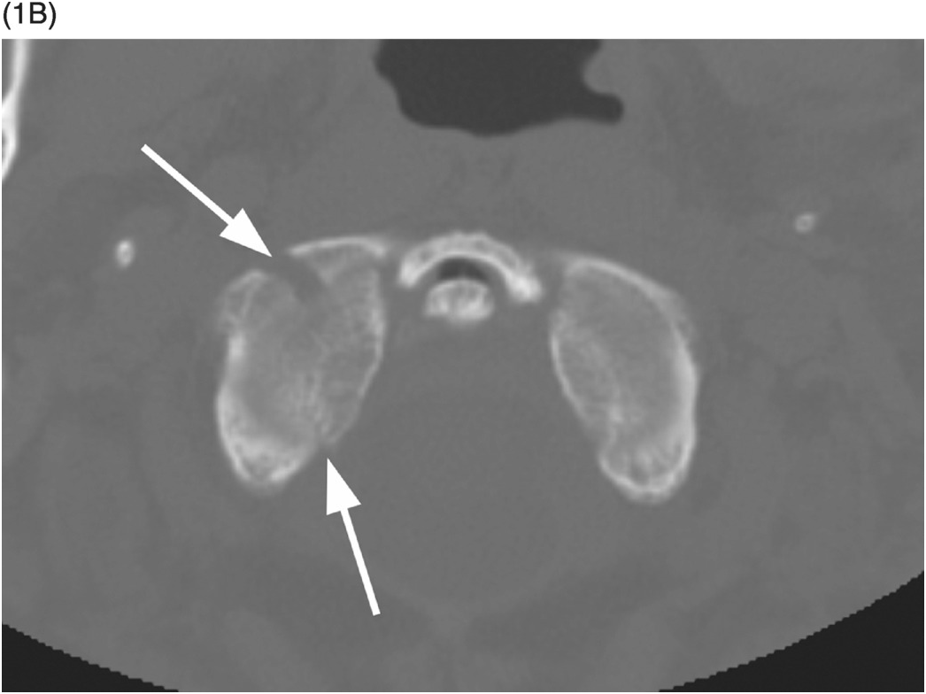
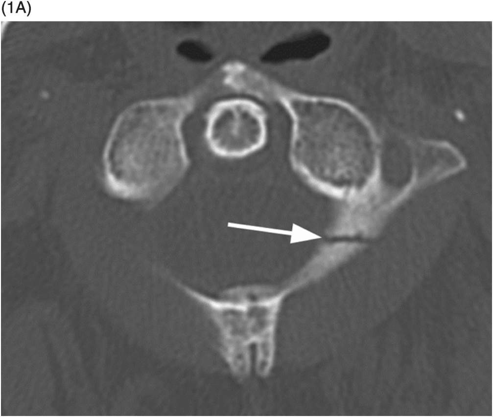
Lucent line through the C1 lateral mass or avulsed bone fragment from the medial portion of the lateral mass, which is best appreciated on an open mouth radiograph and especially CT images, indicate C1 lateral mass fracture. Additional signs are unilateral atlanto-axial displacement or diminished lateral mass height. C1 lateral mass fractures should be fully assessed by CT imaging, where transverse ligament avulsion fracture, intra-articular, or fracture through the transverse foramen should be scrutinized for on coronal and axial images. The fractures may be simple or comminuted and are considered type III atlas fractures.
Differential Diagnosis
Rotational Malalignment
The lateral mass of C1 is displaced medially on C2, by less than 2 mm.
Congenital Clefts of C1 Arches
Well-corticated margins; unilateral or bilateral atlanto-axial offset less than 3 mm.
Clinical Findings, Implications, and Treatment
Integrity of the transverse ligament determines the stability of C1 lateral mass fracture and guides further imaging and treatment. Atlanto-axial lateral displacement greater than 7 mm and atlanto-dental interval greater than 3 mm are indirect signs of transverse ligament rupture recognized on open-mouth and lateral radiographs. Assessed on CT imaging, avulsion at the transverse ligament insertion at the medial part of C1 lateral mass and comminuted lateral mass fracture indicate Dickman type II transverse ligament lesion. Transverse ligament rupture can be well appreciated on axial T1 and T2 MR images as hypointense ligament band disruption. Furthermore, MR is crucial in assessing possible spinal cord injuries by dislocated bone fragments, and is indicated in patients with neurologic deficit. Stable fractures can be treated with rigid cervical collar for 8–12 weeks, while comminuted or unstable fractures are treated either with a rigid cervical orthosis or a halo vest.
Additional Information
Fractures of C1 lateral masses are the least common of all atlas fractures, and occur in flexion injuries with asymmetric axial loading. Fracture extending through transverse foramen should prompt further evaluation of vertebral artery injury, which can cause ischemic events of the posterior circulation and, in more serious cases, occlusion of the basilar artery with death and locked-in syndrome. A unilateral sagittal split subtype of the lateral mass fracture is a rare condition that can cause a subluxation of the occipital condyle into the fracture gap, resulting in late deformity and pain. This type of unusual fracture requires surgical treatment in spite of intact transverse ligament. Intra-articular lateral mass fracture can be the cause of posttraumatic arthritis, associated with chronic occipital neck pain and stiffness as well as neuralgia from intraforaminal compression of C2 nerve.
References
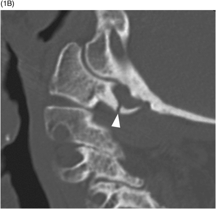
B) Sagittal CT image also demonstrates the nondisplaced fracture line. The ring of the atlas was otherwise intact, without additinal fractures.
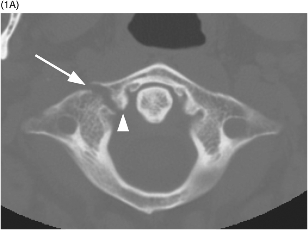
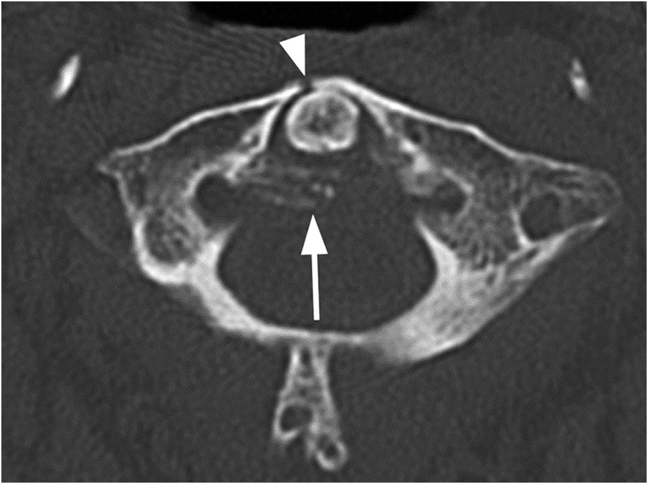
Figure 19.2 Axial CT image in an 82-year-old patient presenting with neck pain following trauma demonstrates an isolated fracture (arrowhead) through the anterior arch of the atlas. Note the otherwise intact ring of C1 and degenerative changes, with calcifications along the transverse ligament (arrow).
Imaging Findings
These injuries are classified as Type I fractures of the atlas. Fracture of the posterior arch is seen on lateral radiographs as a lucent, noncorticated line through the posterior element. It occurs most often near the junction with lateral masses, and is best appreciated on axial CT images due to its most common vertical orientation. Horizontal fracture of the anterior arch is detected on lateral radiograph as a lucent, irregular, noncorticated line and is best appreciated on sagittal CT images. Vertical anterior arch fracture or a “plough fracture” can be occult on radiography, and the comminuted, anteriorly displaced bone fragments are best seen on axial CT images. Retropharyngeal space width of more than 5 mm at the level of C3 body is the indirect sign of anterior arch fracture on lateral radiographs. CT is necessary for complete assessment of these injuries, especially in young children, as atlas may not be visualized on radiographs. Possible associated fractures, primarily of the odontoid process, need to be excluded.
Differential Diagnosis
Posterior Arch Cleft
Sclerotic margins of the bone defect, commonly in the midline.
Anterior Arch Non-United Secondary Ossification Center or Calcification of the Longus Colli Muscle
Well-delineated bone fragment adjacent to the lower pole of the anterior arch without prevertebral tissue swelling.
Clinical Findings, Implications, and Treatment
Isolated fractures of the atlas arches are stable, presenting with minor symptoms such as neck pain and stiffness, dizziness, or headache. However, they are often associated with other cervical spine fractures. Integrity of the transverse ligament is the most important determinant of fracture stability that guides further imaging and treatment. Atlanto-axial offset greater than 7 mm evaluated on open-mouth radiographs and coronal CT images, and atlanto-dental interval (ADI) greater than 3 mm, measured on lateral radiographs and sagittal CT reconstructions, indicate transverse ligament rupture. Ligament injury can be visualized on axial T1- and T2-weighted MR images as a disruption of the hypointense band of the transverse ligament. Stable fractures are treated with rigid collar immobilization for 8–12 weeks, while unstable fractures require either a halo vest or definitive surgical treatment.
Additional Information
Posterior arch fracture occurs during hyperextension, when the junction with lateral masses is compressed between the occiput and the spinous process of C2. Horizontal anterior arch fracture is an avulsion fracture at the insertion of the longus colli muscle or anterior atlanto-dental ligament. “Plough fracture” is a serious, although extremely rare, condition, occurring during hyperextension, when the anterior arch is shorn off, anteriorly displaced, and fragmented by the odontoid process. It is unstable and can be accompanied by atlanto-occipital dissociation and posterior displacement of the cranium relative to the cervical spine.
References
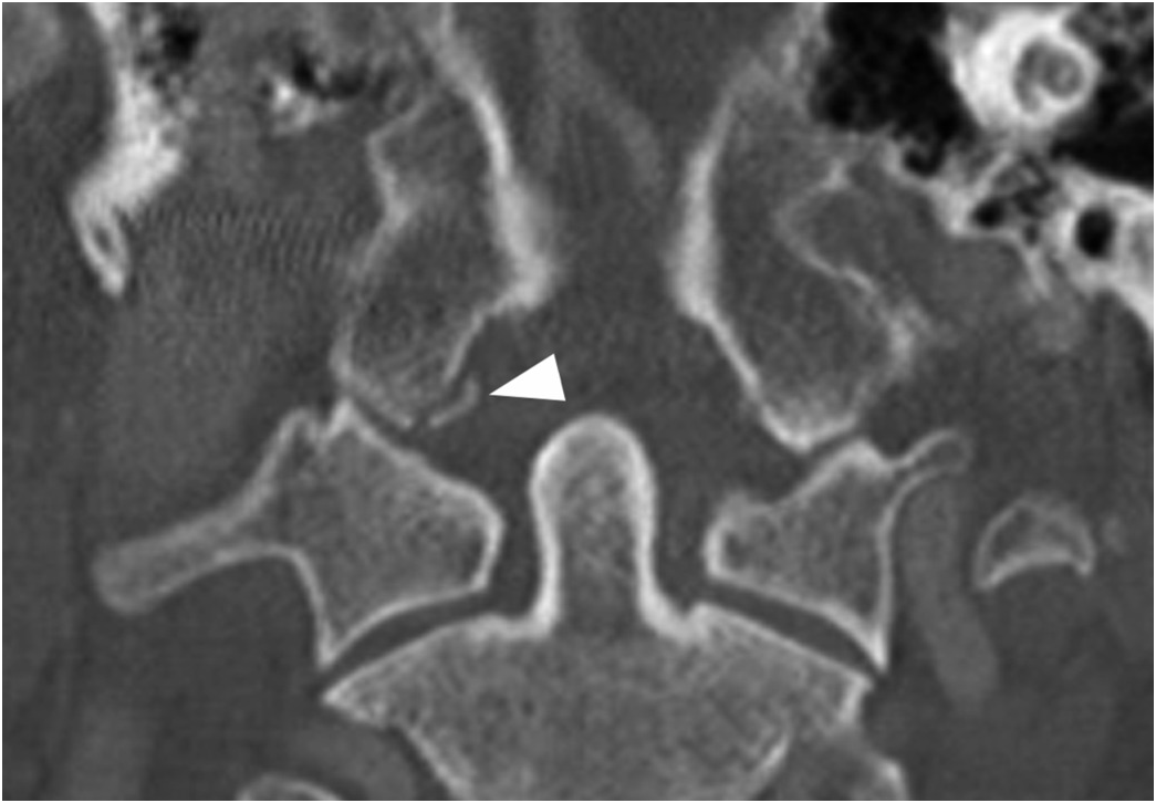
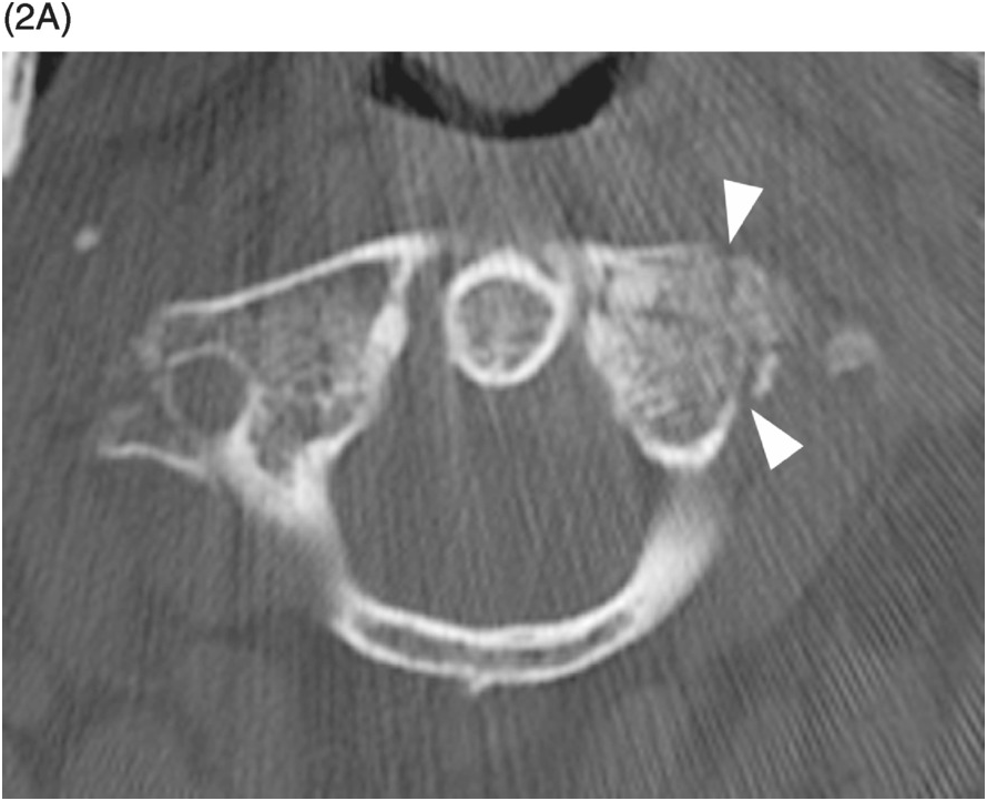
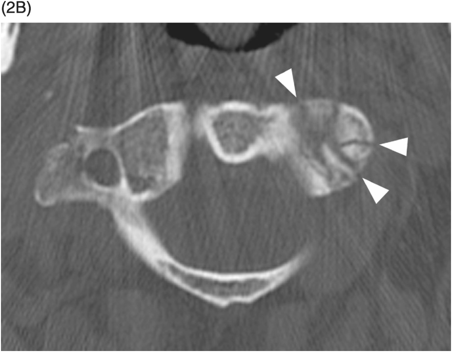
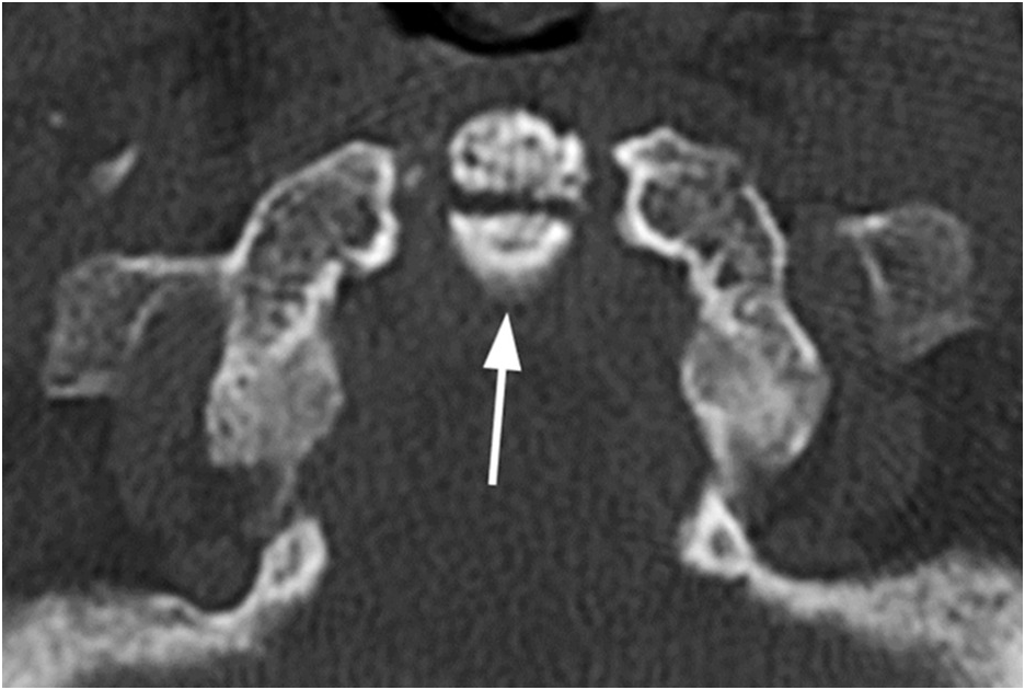
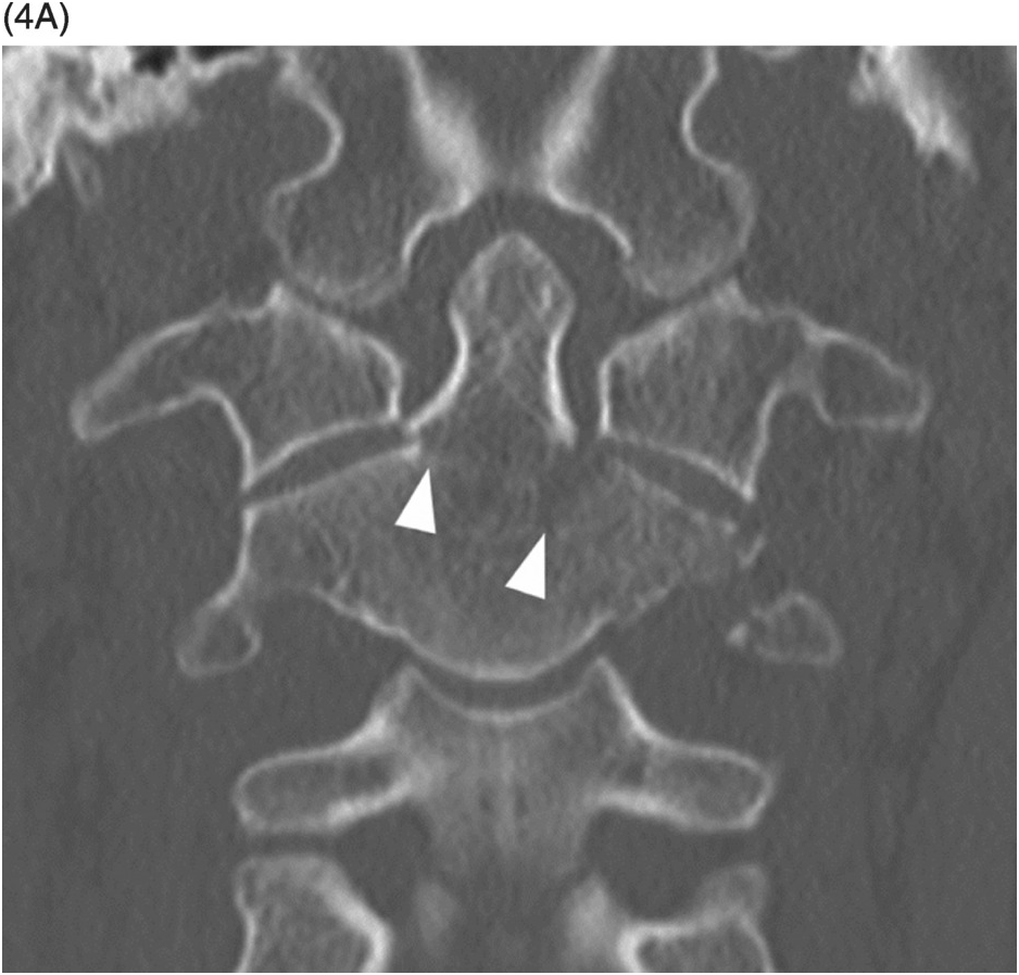
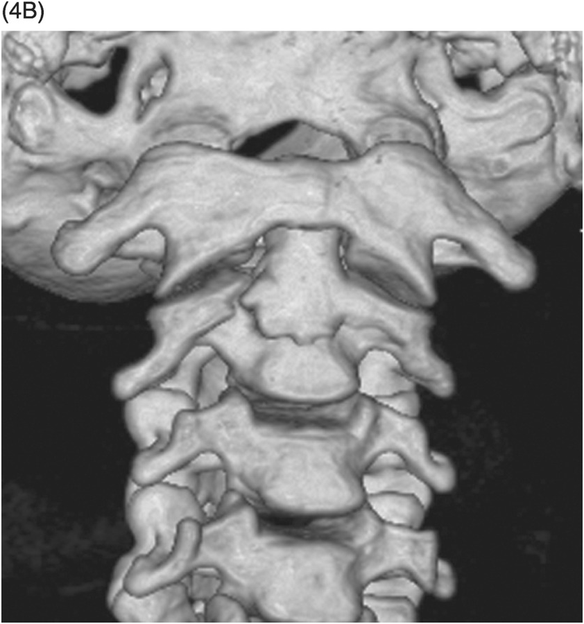
Figure 20.4 CT of an unstable type 3 odontoid fracture in a different patient following a fall. The fracture lines (arrowheads) are extending into the body of C2 and left lateral mass with fragment displacement on coronal (A) and 3D volume rendered (B) images.
Imaging Findings
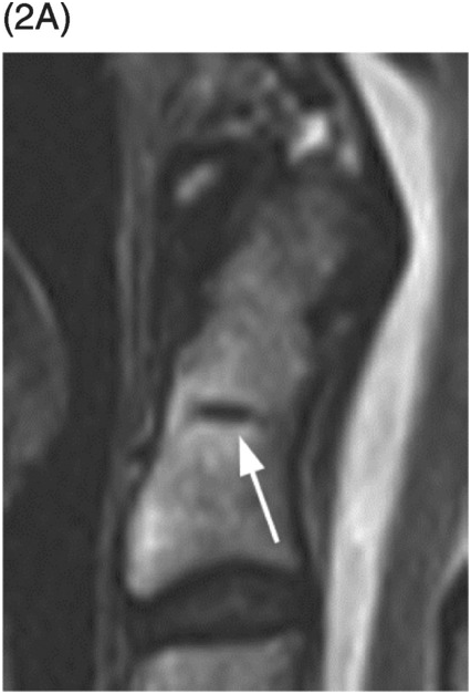
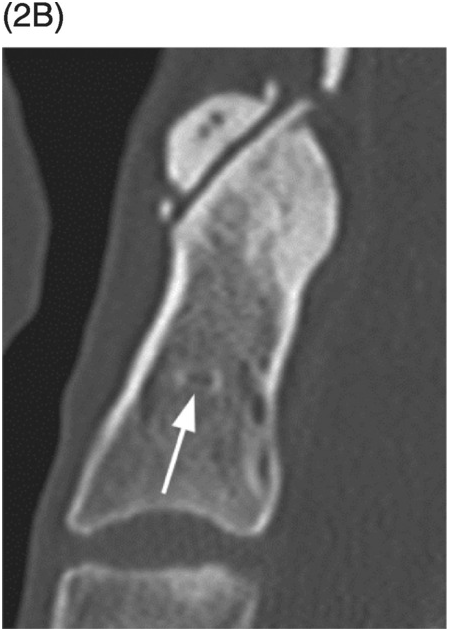
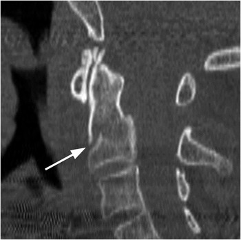
Odontoid fracture type 1 is an avulsion fracture of the odontoid tip. It is recognized as a very small bony fragment with irregular and noncorticated margins avulsed from the lateral side of the odontoid tip at the alar ligament attachment. Type 3 odontoid fracture extends from the dens through the body and/or lateral masses of C2 vertebra. Widened prevertebral tissue (>6 mm at C2 level) and disruption of the axis or ‘‘Harris’’ ring on lateral radiographs are the signs of type 3 fracture. Dentocentral synchondrosis remnant is an important anatomical landmark for distinguishing type 2 from type 3 odontoid fractures. It represents the border between the base of the odontoid process and the body of C2 vertebra and is situated below the superior border of C2 lateral masses. It can be seen in the majority of patients as a hypodense horizontal line on sagittal T1- and T2-weighted images, or less commonly as a sclerotic line on sagittal CT images. Fracture orientation angle, fragment dislocation, angulation and comminution, as well as possible additional craniocervical junction injuries should be searched for and evaluated on reformatted CT images to assess fracture stability. Injury to the alar and transverse ligaments, tectal membrane, prevertebral tissues, presence of epidural hematoma, pseudomeningocele, and spinal cord injury are assessed on MRI.
Differential Diagnosis
Persistent Ossiculum Terminale
Nonfused secondary ossification center along the tip of the odontoid process, small bone fragment with smooth corticated margins of disproportionate shape and size to the V-shaped cartilaginous cleft along the subjacent superior margin of the odontoid process.
Os Odontoideum
Well-corticated bone fragment along the superior margin of the odontoid process accompanied by a foreshortened base of the dens and hypertrophied anterior C1 arch.
Clinical Findings, Implications, and Treatment
Odontoid fractures result from both hyperflexion and hyperextension injuries to the cervical spine. They typically occur in young patients involved in a high-energy trauma. In the elderly with osteopenic bone, even a low-energy trauma can fracture the odontoid. Cervical spine CT is recommended as the initial imaging modality due to low sensitivity and specificity of radiography and often very subtle clinical findings. MRI is indicated in patients whose neurological status cannot be assessed and in those with suspected spinal cord or nerve root injury. Type 1 fractures are avulsion fractures involving the apical and alar ligaments. These fractures are stable when at least one alar and the transverse ligament are intact. Most type 1 odontoid fractures are stable and heal well with cervical immobilization. In extreme cases with bilateral alar ligament injury, type 1 odontoid fracture may be associated with atlanto-occipital instability that requires occipitocervical fusion. Type 3 odontoid fractures heal well with cervical immobilization because of large bony contact area and adequate vascular supply. In nondisplaced type 3 fractures, rate of healing, with cervical immobilization, is close to 95%. Risk factors for non-union include fractures that involve the waist of the dens or when there is anterior or posterior displacement. Vertically displaced type 3 fractures are extremely unstable and associated with brainstem and spinal cord injury. Anterior fixation may render best results in younger patients, while posterior spinal fusion may be the treatment of choice in the elderly.
Additional Information
Odontoid fractures in the elderly deserve separate mention because of higher associated mortality and morbidity. Reasons include higher incidence of osteopenia, associated comorbidities, and poor rehabilitation potential. In-hospital mortality can be close to 35% in patients aged >70 years. These patients also tolerate halo-vest immobilization poorly because of impaired cardiopulmonary reserve. Hence many surgeons recommend early surgical treatment of odontoid fractures in the elderly when there is significant risk of non-union.
References
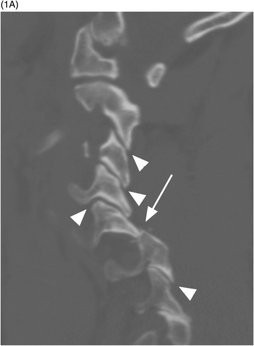
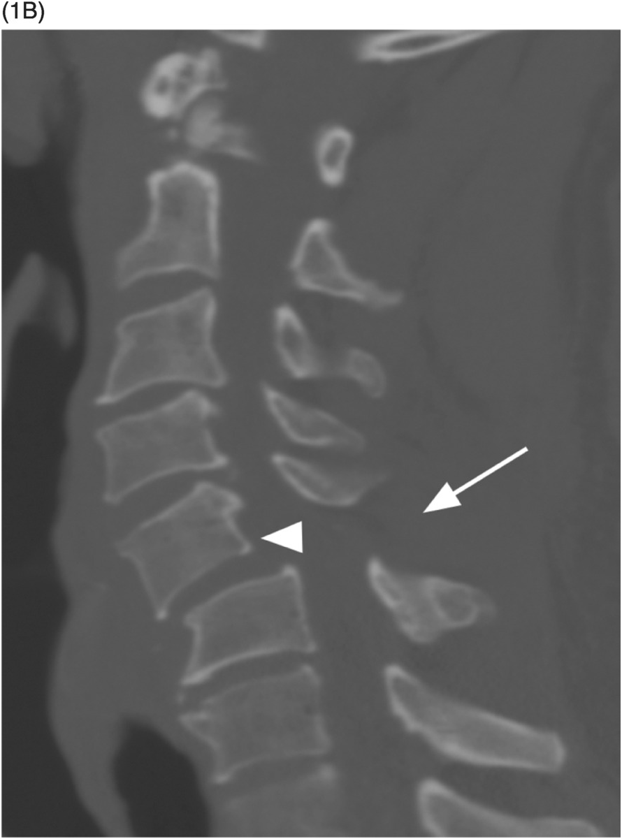
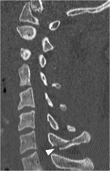
B) Mediosagittal CT image demonstrates C5 anterolisthesis (arrowhead) and widening of the C5–C6 interspinous and intralaminar space (arrow).
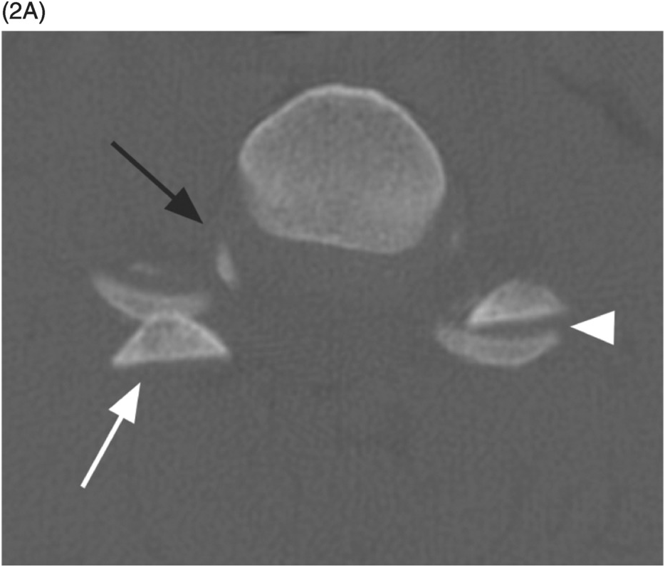
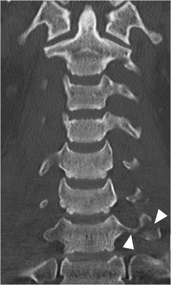
A) Axial CT image in a different patient reveals the “reverse hamburger” sign on the right (white arrow) with opposing convex cortical surfaces, consistent with unilateral locked facets. Note the normal relationship of the adjacent flat articular surfaces on the left side (arrowhead), resembling a hamburger bun. There is also malalignment of the right uncovertebral joint (black arrow) with rotation and widening of the joint space.
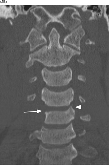
B) Coronal CT shows misalignment of the right C4–C5 uncovertebral joint (arrow). The uncovertebral process appears to be missing on this image. Compare to the normal contralateral (arrowhead) and bilateral uncovertebral joints at other levels.
Imaging Findings
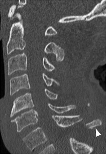
Axial CT images show unilateral “reverse hamburger” (originally “reverse hamburger bun”) sign with the convex nonarticular surfaces facing each other, in contrast to the normal approximation of flat articular surfaces. Ipsilateral uncovertebral malalignment is another reliable sign on axial or coronal images. Midsagittal CT images demonstrate anterolisthesis of the superior vertebra along with widening of the interlaminar and interspinous space. More lateral CT images are the key, as they directly show the locked facets, in contrast to the normal alignment of other ipsi- and contralateral facet joints (similar to shingling on a roof). Associated small fracture fragments may also be present. Spinous processes of the involved vertebra and those above are rotated toward the side of the lock. The images should be scrutinized for fractures involving the adjacent pedicles, laminae, and articular pillars. MRI may reveal associated injuries to the ligaments, spinal cord, and vertebral artery.
Differential Diagnosis
Bilateral Facet Dislocation
Locked facets are present on both sides.
Fractured Articular Pillar (Facet Fracture)
Small fracture fragments are frequently found with unilateral facet lock; however, the fracture does not extend through the entire articular pillar.
Flexion Sprain
Widening of the interspinous space with possible mild subluxation but without facet joint dislocation.
Clinical Findings, Implications, and Treatment
These patients typically present with neck pain, sometimes associated with radiculopathy, while spinal cord injury is uncommon. The injuries are mostly stable but require prompt stabilization and treatment. Closed traction-reduction followed by anterior fusion (ACDF) or posterior instrumented fusion (with or without preoperative closed traction-reduction) is an effective treatment. Combined anterior and posterior surgical approach with fusion is commonly performed in patients with spinal cord injury. Following appropriate treatment, neurological deficits improve or resolve in a majority of affected individuals.
Additional Information
The mechanism of this injury is a combination of lateral flexion and distraction. Although facet dislocation most commonly occurs in the lower cervical region, it may affect any spinal level, all the way to the lumbosacral junction. Patients with cervical facet dislocation and spinal cord injury (SCI) tend to present with a more severe degree of initial trauma and display less potential for motor recovery, compared to SCI patients without facet dislocation. Facet dislocations without fractures have a significantly higher association with cord syndromes than do facet injuries with fractures. A rare case of unilateral facet dislocation as a preexisting lesion, possibly originating from a forceps delivery at birth, has been described, with full clinical recovery following conservative approach.
References
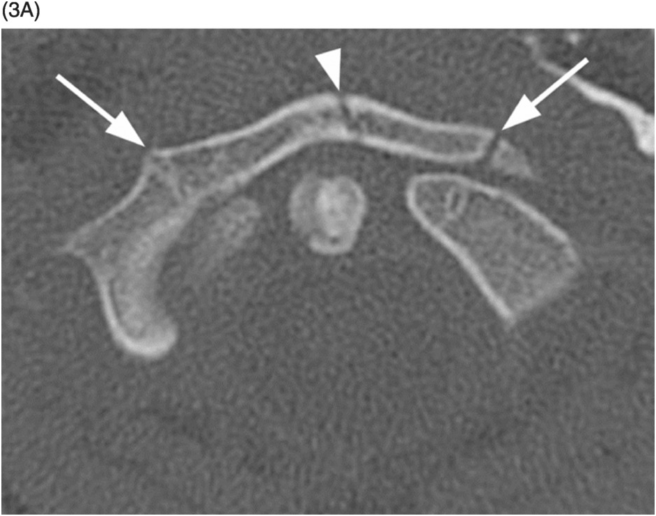
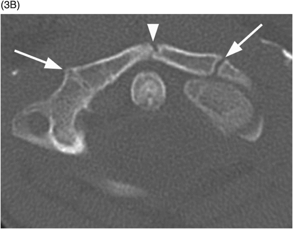
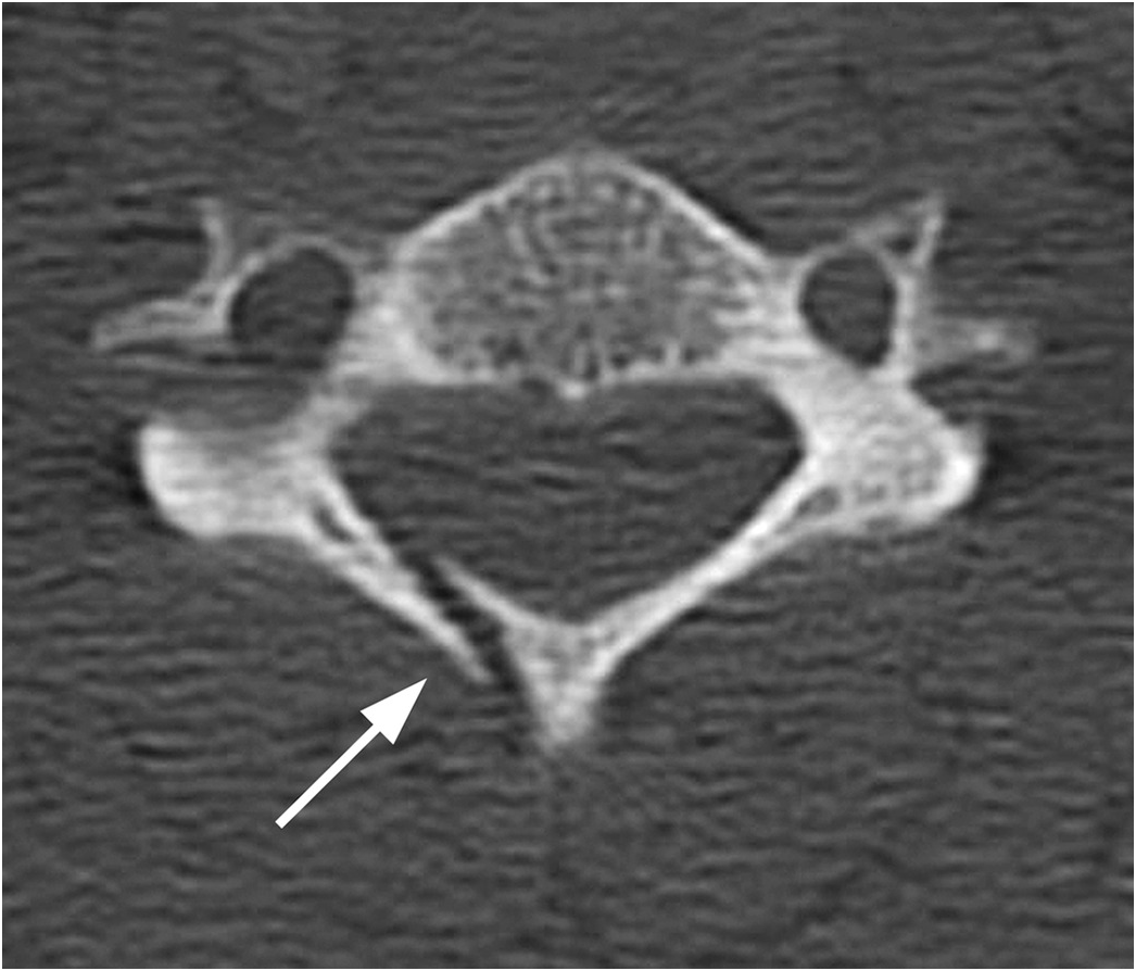
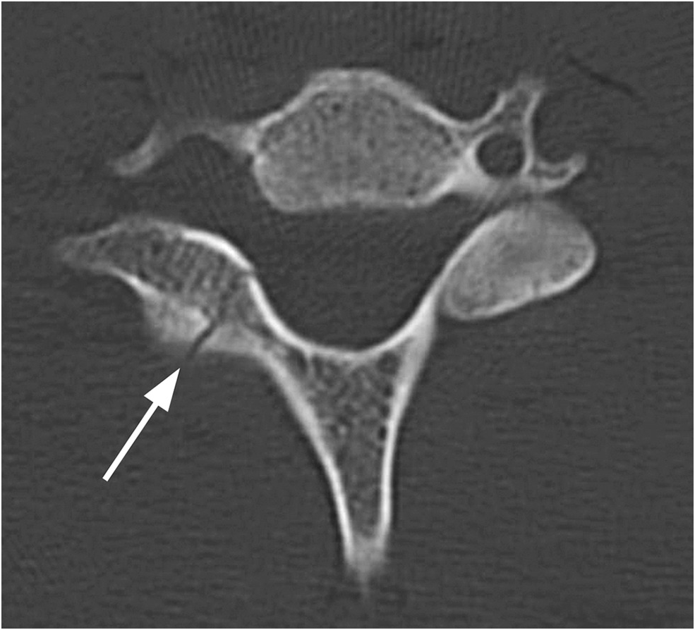
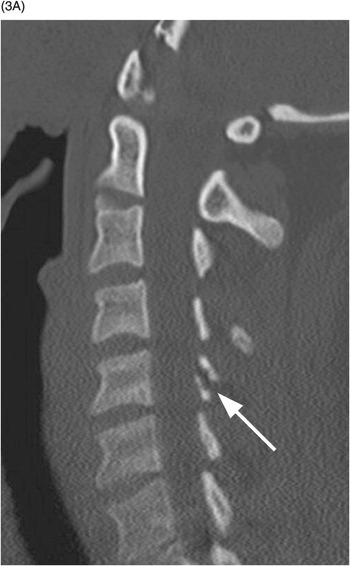
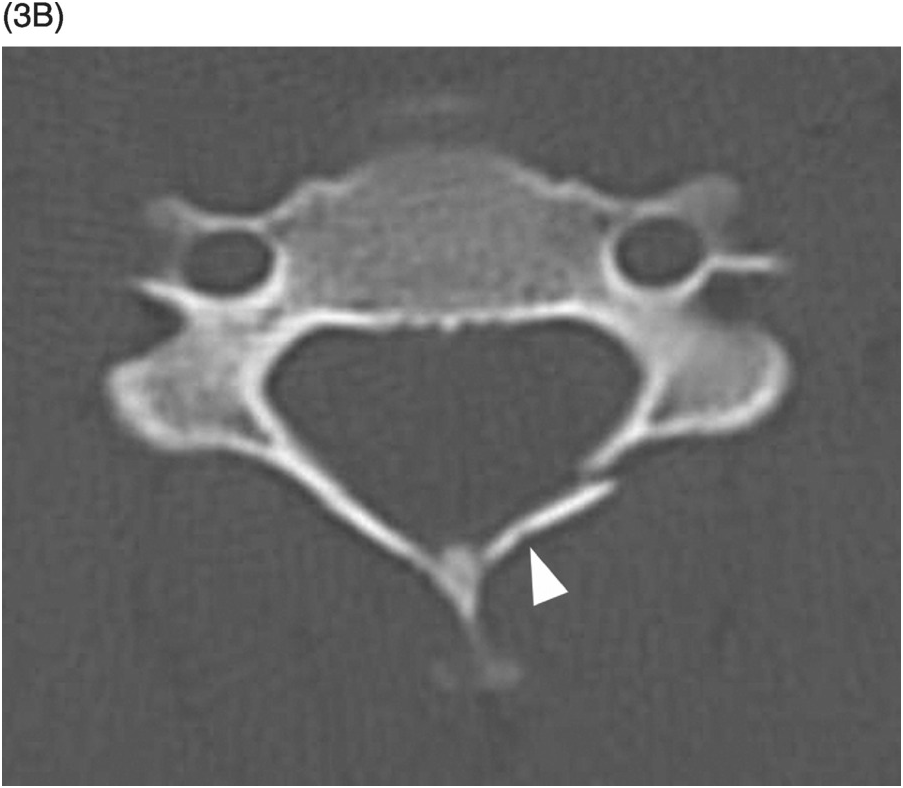
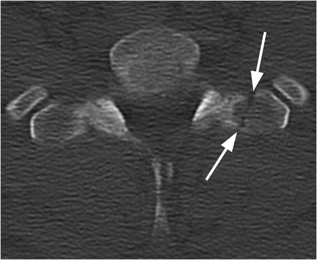
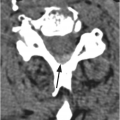
B) Axial CT image shows that there are actually two fracture lines: a complete one more anteriorly and a greenstick type more posteriorly (arrowhead). There were no additional fractures in this patient.
Imaging Findings
Radiographs show disruption of the spinolaminar line, while CT directly reveals fracture of the lamina, which is usually best seen in the axial plane. Laminae lesion may be complete or of the greenstick type (incomplete, the fracture line does not extend through the entire thickness of the bone). The fracture line may extend to the spinous process and/or contralateral lamina, articular pillar/facet joint, and transverse process/transverse foramen, which should be carefully searched for as the presence of those lesions excludes an isolated laminar fracture and is indicative of a more serious injury. Depressed fractures of the lamina may be associated with spinal cord injury.
Differential Diagnosis
Burst Fracture with Laminar Fracture
Obvious additional fracture(s) with compression of the vertebral body and retropulsion of fragment(s) into the spinal canal, frequent associated ligamentous injury.
Clinical Findings, Implications, and Treatment
Isolated fractures of the lamina have been described in all age groups, including infants. These fractures usually occur after a hyperextension injury, and the patients typically present without any neurologic compromise. Isolated fracture of the lamina caused by direct trauma to the posterior neck may, however, lead to a significant neurologic deficit, which needs to be treated surgically with posterior decompression.
Additional Information
Isolated laminar fractures, typically involving the cervical spine, are often missed on radiographs and had been considered very uncommon. However, with modern CT scanners, these lesions are now documented more frequently.
On the other hand, laminar fractures are commonly associated with burst fractures. In upper lumbar burst fractures, complete lamina fracture is an indicator of injury severity, frequently accompanied by dural tears and nerve root entrapment. Open-book laminectomy has been therefore recommended, even if the patient is neurologically intact.
“Spinolaminar breach,” or disruption of the spinolaminar line, is a sign on plain films, which indicates a complex spinous process fracture with extension into the lamina and spinal canal. Misdiagnosis of a “clay shoveler’s fracture” should be avoided, as spinous process fractures with spinolaminar breach may have associated posterior ligamentous injury, which carry a significant potential for delayed instability and neurological deficit.
References
Imaging Findings
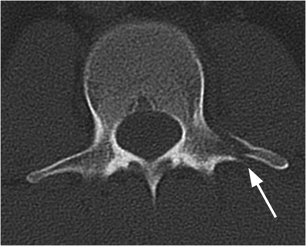
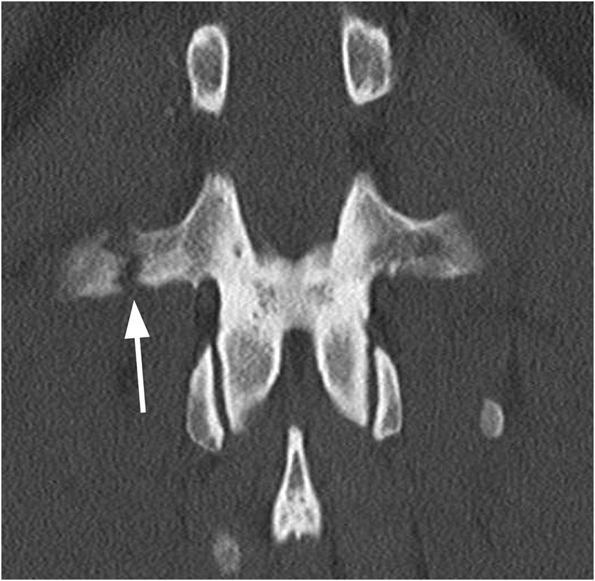
CT is the modality of choice for detection of these fractures, as up to 60% of lumbar transverse process fractures identified on CT will be missed on plain radiographs, primarily the nondisplaced ones. CT readily identifies these fractures, even on just axial images, as lucent lines without sclerotic margins, usually with irregular jagged edges. These injuries most frequently occur in the upper lumbar spine and are commonly multiple. Fractures of transverse processes in the thoracic and lumbar spine are clinically insignificant on their own, but they are associated with structurally unstable thoracolumbar spine fractures in 10–20% of patients. Plain radiographs miss these additional unstable thoracolumbar bony injuries in about 10% of cases. Upper thoracic and L5 fractures may be associated with rib and pelvic fractures, respectively. Also, transverse process fractures typically occur with high-energy blunt traumas, often resulting in associated injury of other organs, which should be carefully searched for.
Fracture of transverse process in the cervical spine is of special significance, as the fracture may extend into the transverse foramina, indicating possible vertebral artery injury. Cervical transverse process fractures also have a strong association with other cervical spine fractures and blunt cerebrovascular injury.
Differential Diagnosis
Secondary Ossification Center
Smooth corticated margins, at the tip of the transverse process with a somewhat rounded appearance.
Chronic Transverse Process Fracture
Sclerotic margins, with a smooth appearance instead of at least somewhat irregular edges.
Clinical Findings, Implications, and Treatment
The most common mechanism of injury is motor vehicle accident (MVA), followed by falls. MVAs appear to be more commonly the cause in pediatric patients, while falls are more frequent cause in adults. Patients with isolated transverse process fractures (not involving the transverse foramina in the cervical spine) do not present with neurological deficit.
These fractures can be treated conservatively without concern for long-term outcome sequelae such as pain, neurologic deficits, or ambulatory difficulties. Management strategies involve unrestricted movement, bracing, and orthotics.
Additional Information
Acute traumatic isolated transverse process fractures are increasingly identified in traumatized patients due to the increased routine use of CT in trauma. While theses lesions are common sequelae of trauma and considered a minor and stable fracture in the lumbar and thoracic spine, there is strong association between these fractures and other traumatic injuries. Approximately 10–50% of patients with lumbar transverse process fractures have hepatic and splenic as well as genitourinary and diaphragmatic injuries.
References
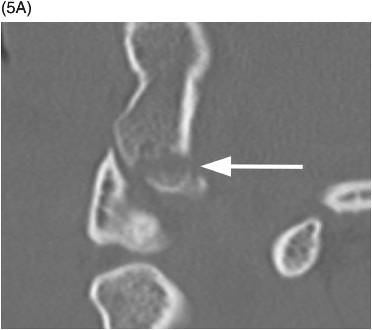
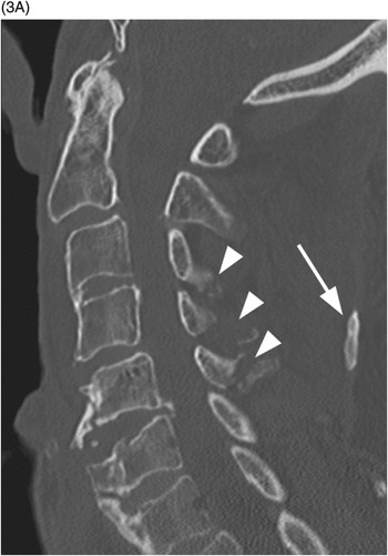
Figure 24.2 Midsagittal CT image shows C7 spinous process fracture with displaced distal fragment (arrowhead).
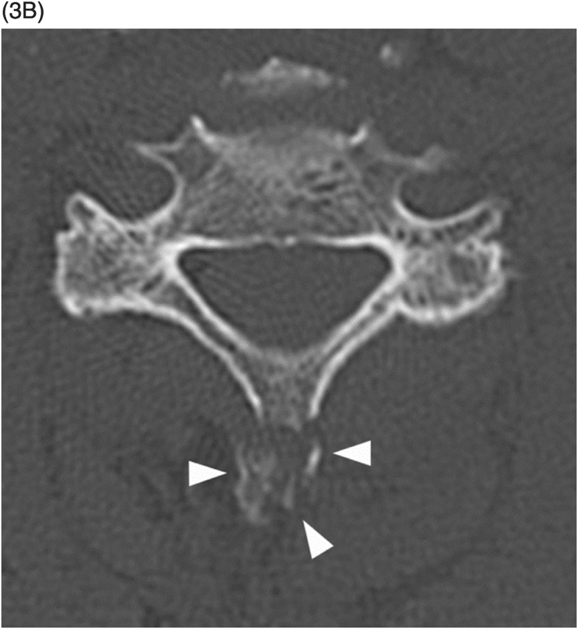
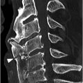
A) Sagittal CT image shows multiple spinous process fractures, which may be an indicator of more severe injuries. Anteriorly increased C5–C6 disc space and a probable fracture of the osteophyte are suggestive of hyperextension and anterior longitudinal ligament injury. Note ossification of the nuchal ligament (arrow).
B) Axial CT image at C5 level reveals that the spinous process fracture is comminuted (arrowheads).
Imaging Findings
Isolated fractures of the spinous process most commonly occur in the lower cervical and upper thoracic spine. A double spinous process shadow is a sign of spinous process fracture on anteroposterior radiographs, while a downward displacement of a bony fragment or a breach of the spinolaminar line can be detected on lateral x-rays. Nevertheless, a spinous process fracture can be missed on plain films, primarily nondisplaced fractures and those that are superimposed over ribs. CT is more sensitive for diagnosing bone fractures, especially small bone fragments. MRI enables precise evaluation of bone marrow and soft tissue, including ligamentous injury.
Differential Diagnosis
Pseudoarthorsis Due to a Non-Union Spinous Process Fracture
Bone fragment with well-corticated margins adjacent to the spinous process, no signs of bone marrow edema, ligamentous or paraspinal soft tissue injury on MRI.
Clinical Findings, Implications, and Treatment
Injury mechanism of the isolated spinous process fracture can be an avulsion due to increased shear forces of the attached ligaments and muscles, direct blow to the flexed neck or back, hyperflexion or hyprextension injury, and a stress fracture, recently more commonly recognized in weightlifters, golfers, jockeys, and other athletes. Patients typically present with neck or back pain without neurologic deficits, and severe tenderness with or without crepitation over the tips of the affected spinous processes. History of significant traumatic event may be negative, especially in cases of stress fractures; however, a typical clinical presentation should raise suspicion and prompt further imaging. Spinous process fractures are stable and do not cause any neurological damage on its own. However, multiple fractures might be an indicator of more severe injuries to the spine or surrounding tissues and organs. Isolated spinous process fractures are treated conservatively with immobilization and restriction of physical activity for 4–6 weeks. Treatment results are very good in terms of pain relief and range of motion despite a high incidence of non-union. Pseudoarthrosis develops often and is usually asymptomatic. However, if pain associated with pseudoarthrosis persists longer than 10 weeks, surgical removal of the non-united bone fragment is indicated.
Additional Information
Isolated fractures of the lower cervical and upper thoracic spine were first described in 1933 as “clay shoveler’s fracture” among hard-labor workers in Australia, who tossed clay from the ditches with long-handled shovels over their shoulders. The clay sometimes stuck to the shovel, producing a sudden, unexpected opposite force to the neck and back muscles, causing an avulsion fracture at the attachment site on the spinous processes.
References

