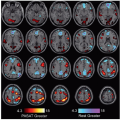
Sentence Completion
Lubdha M. Shah, MD
Key Facts
Terminology
Visual language comprehension task for language localization
Anatomy-based Imaging Issues
Language localization in Broca area (inferior frontal gyrus) and Wernicke area (posterior superior temporal sulcus) and hippocampal structures
Eye movement and attentional areas (precentral sulci, medial frontal lobes, intraparietal sulci)
Working memory (middle frontal gyri)
Primary and associative visual areas (occipital areas)
Timing
Block design: 4 minutes
Instructions
Subject is shown sentences that end in a blank
Subject reads them silently and thinks of a word to fill in the blank (e.g., “They put the dirty dishes in the __________”)
Control task: Rest with eyes open
Visual fixation point
Preprocessing
Distortion, motion, slice timing correction
Coregistration to MPRAGE or T2 anatomic image for overlay
Applications
Task elucidates areas important to reading comprehension in posterior and inferior temporal regions that verbal fluency and auditory discrimination tasks do not
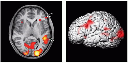
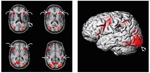 (Left) Axial MPRAGE with BOLD signal overlay demonstrates bilateral expressive speech areas
 as well as receptive speech areas as well as receptive speech areas  . Bilateral speech representation may be related to the left perisylvian encephalomalacia secondary to the prior middle cerebral artery infarction . Bilateral speech representation may be related to the left perisylvian encephalomalacia secondary to the prior middle cerebral artery infarction  . (Right) Sagittal surface rendering in the same patient illustrates inferior frontal gyrus activation . (Right) Sagittal surface rendering in the same patient illustrates inferior frontal gyrus activation  and a small focus of posterior superior temporal gyrus activation and a small focus of posterior superior temporal gyrus activation  . This task elicits marked activation in the visual cortex . This task elicits marked activation in the visual cortex  . .Stay updated, free articles. Join our Telegram channel
Full access? Get Clinical Tree
 Get Clinical Tree app for offline access

|
