Abstract
Hemophilia is a genetic disorder that manifests due to a deficiency of clotting factors. One of the most common complications is hemophilic arthropathy which results in bleeding into large joints causing hemosiderin deposition. Recurrent hemarthrosis causes joint destruction resulting in pain and a decreased range of movements. Radiological imaging is essential for staging, prognostication, and treatment protocol. X-rays show the extent of joint destruction and narrowing of joint space. Ultrasonography helps in visualizing tendons and joint effusion. However magnetic resonance imaging is the gold standard for staging hemophilic arthropathy. Management involves both medical and surgical intervention as per the level of joint involvement.
Background
A lack of coagulation factor VIII causes hemostasis to be compromised in hemophilia A, an X-linked recessive genetic disease [ ]. Hemophilia is divided into hemophilia A and hemophilia B based on the type of coagulation factor the patient lacks. The results of factor VIII and factor IX measurements are used to categorize the disease’s severity [ ]. Hemophilia may manifest in many ways depending on the activity of clotting factors. The long-term result of recurring hemarthrosis is hemophilic arthropathy (HA), a chronic joint disease. About half of hemophiliacs will get severe arthropathy [ ].
Hemophilia is characterized by a tendency to bleed, especially in synovial joints like the elbows, knees, and ankles [ ]. The fourth most commonly reported source of bleeding is the shoulder joint [ ]. A patient’s quality of life can be seriously impaired by shoulder pain and decreased joint movements. The vicious cycle of chronic synovial inflammation and thickening, neo-angiogenesis, rebleeding, remodeling, and finally osteochondral surface damage and joint space narrowing can be initiated by a single episode of hemarthrosis, either clinically overt or undiagnosed subclinical bleeding [ ]. Patients usually present with symptoms of stiffness, redness of surrounding skin, and restriction of the mobility of the joint involved.
In these situations, radiological diagnostic techniques are crucial. It facilitates the division of the joint degenerative process into different stages of pathology and clinical manifestation [ ]. It also helps with orthopedic exploration and radiological scoring systems [ ]. The most often utilized methods are magnetic resonance imaging (MRI), X-rays, and ultrasonography. Despite their drawbacks, X-rays are still the most popular imaging technique. Its limited sensitivity for identifying anomalies in cartilage and synovial membranes is its primary flaw. The gold standard for examining joint alterations in hemophiliac patients is magnetic resonance imaging (MRI). In the early phases of hemophilic arthropathy, it is the most effective instrument for analyzing soft tissues and osteochondral changes. However, its use and periodic follow-ups to ascertain the evolution of acute and chronic processes are limited by its high prices, limited accessibility, evaluation time, and challenging applicability in youngsters [ ]. The Denver scale and the European scale are 2 parameters used in MRI joint injury assessment.
Compressive bandages, analgesics, and the administration of bleeding factors are typically used as treatments. However, depending on the kind of joint and degree of damage, surgical intervention, such as synovectomy, joint debridement, arthrodesis, and arthroplasty, is frequently necessary when nonsurgical therapy fails in patients with chronic arthropathy progression [ ]. In cases of severe end-stage degenerative joint degeneration, joint arthroplasty is recommended.
Case presentation
We report the case of a 40-year-old male who is a known case of hemophilia A and presented with complaints of pain in his right shoulder for 2 months. He was unable to abduct his right shoulder. The patient initially experienced dull aching, persistent pain which was associated with swelling in his right shoulder. His symptoms were progressive. His range of movements in his right shoulder was decreased and was painful. He was initially able to do his daily activities but now due to decreased movements and excessive pain, he was not able to perform his daily activities like shaving or dressing. On admission, his vitals were stable with a pulse of 82 beats per minute, blood pressure of 110/70 mm Hg, and respiratory rate of 16 breaths per minute. He had a history of recurrent bleeding episodes following minor trauma. He had been transfused blood multiple times in the past. He had no history of major accidents, drug allergies, or any infectious disease. The patient’s mother is a known case of hemophilia A and has a history of recurrent nasal bleeds and easy bruising. Three years back patient had a similar episode of inflammation and pain in the left knee which was diagnosed as hemarthrosis of the knee joint which caused a pathological fracture in his left femur shaft and was operated upon. An interlocking nail was used to fix the fracture. The patient had no previous surgical interventions. The patient also complained of new-onset pain in his right knee but there was no restriction of mobility. Laboratory investigations are shown in Table 1 .
| Parameters | Values | Normal range |
|---|---|---|
| Hemoglobin | 9.8 gm% | 13-17 gm% |
| Total leukocyte count | 6500 cells/cu mm | 4,000-10,000 cells/cu mm |
| Total platelet count | 1.76 lakh/cu mm | 1.5-4.1 lakh/cu mm |
| Mean corpuscular volume | 71.1 fL | 83-101 fL |
| Urea | 38 mg/dl | 19-43 mg/dl |
| Creatinine | 0.9 mg/dl | 0.2-1.3 mg/dl |
| Serum Sodium | 137 mm/L | 135-145 mm/L |
| Serum Potassium | 3.9 mm/L | 3.5-5.1 mm/L |
| INR | 1.5 | 1-1.3 |
| Alkaline phosphatase | 62 U/L | 38-126 U/L |
| Alanine aminotransferases | 33 U/L | <50 U/L |
| Aspartate aminotransferase | 44 U/L | 17-59 U/L |
| Albumin | 3.9 g/dL | 3.5-5 g/dL |
| Total bilirubin | 1.2 mg/dl | 0.2-1.3 mg/dl |
| Magnesium | 1.9 mg/dl | 1.6-2.2 mg/dl |
| Phosphorous | 2.1 mg/dl | 2.5-4.5 mg/dL |
| Calcium | 7.7mg/dl | 8.4-10.2 mg/dl |
The patient underwent radiological evaluation for the condition of his various joints. X-ray anteroposterior view of his left knee joint showed an interlocking nail in situ. X-ray anteroposterior bilateral knee showed changes of osteoarthritis (left>right) in the form of marginal osteophytes, loss of joint space, and subchondral sclerosis. There was a widening of the inter-condylar notch and flattened condylar surface ( Fig. 1 ).

Related posts:
 A rare and life-threatening case of spontaneous hemopneumothorax presenting with severe hemodynamic instability
 A very rare case of ileocolic and appendiceal intussusception with acute appendicitis
 Hepatic and extra-hepatic hydatid cysts: A case series of radiological and clinical insights
 Mid-term follow-up of a fractured flow diverter in the internal carotid artery
 An atypical multiple autoimmune syndrome: A case report including myocarditis
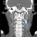 Persistence of the hypoglossal artery: A rare case report
Stay updated, free articles. Join our Telegram channel

Full access? Get Clinical Tree


