Silent Verb Generation
Lubdha M. Shah, MD
Key Facts
Anatomy-based Imaging Issues
Fluency-verb generation task produces consistent activation of both frontal and posterior language areas
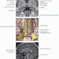
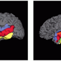
Broca area classically located in pars opercularis and posterior portion of the pars triangularis of the inferior frontal gyrus
Wernicke area involves portions of supramarginal gyrus, angular gyrus, posterior superior and middle temporal gyri, planum temporale
Design
Block design: 4 minutes
Verbal fluency paradigms primarily require language expression and secondarily require language comprehension
Activation in classic Broca area & often in Wernicke area in the dominant hemisphere
Silent word production reduces motion artifacts significantly compared with overt word production
However, task performance cannot be monitored
Instructions
Subject is asked to generate a verb from a presented noun or picture
Applications
Proximity of a lesion to the functional language areas can be assessed
Images can be used by neurosurgery for pre- and intraoperative surgical planning
Assessment of hemispheric dominance
Pitfalls
Between-session variance mostly due to variability of underlying cognitive processes & motion correlated with stimuli
Not due to technical limitations of data processing
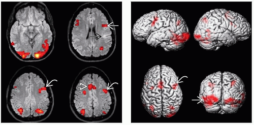 (Left) Axial FLAIR MRI with BOLD signal overlay demonstrates activation in left inferior frontal gyrus (IFG)
 and dorsolateral prefrontal cortex (DLPFC) and dorsolateral prefrontal cortex (DLPFC)  during silent verb-generation task. The IFG lies anterior to the abnormal cortical hyperintensity during silent verb-generation task. The IFG lies anterior to the abnormal cortical hyperintensity  . There is also activation in the supplementary motor area . There is also activation in the supplementary motor area  . (Right) 3D surface rendering (same patient) shows activation in left IFG in the expected expressive speech area . (Right) 3D surface rendering (same patient) shows activation in left IFG in the expected expressive speech area  . Note bilateral occipital lobe activation . Note bilateral occipital lobe activation  in the expected visual cortices. in the expected visual cortices.Stay updated, free articles. Join our Telegram channel
Full access? Get Clinical Tree
 Get Clinical Tree app for offline access

|
