KEY FACTS
Terminology
- •
Benign, fluid-filled, nonneoplastic renal lesion
Imaging
- •
Ultrasound is ideal for characterizing simple or complex renal cysts in nonobese patients
- •
Unilocular, thin-walled, round/oval renal lesion
- •
Anechoic: No internal echoes, septations, or solid components; distinct posterior wall
- •
Increased sound transmission gives rise to characteristic posterior acoustic enhancement
- •
Variable size
- •
Location: Intraparenchymal, exophytic, parapelvic
Top Differential Diagnoses
- •
Complex renal cyst
- •
Peripelvic cysts
- •
Prominent pyramids
- •
Cystic disease of dialysis
- •
Perinephric collections
- •
Pyelogenic cyst/pyelocalyceal diverticulum
- •
Multilocular cystic nephroma
Clinical Issues
- •
In 20-30% of middle-aged adults and 50% > 50 years of age
- •
Most common renal lesion, usually detected incidentally
- •
No further imaging or monitoring of cyst is warranted
- •
May present with pain from bleeding/rupture/infection or mass effect when large
- •
Multiple renal cysts may indicate syndrome, such as autosomal dominant polycystic kidney disease
Diagnostic Checklist
- •
Well-defined, round or ovoid renal lesion with posterior acoustic enhancement, distinct echogenic posterior wall, and complete lack of internal echoes (anechoic)
Scanning Tips
- •
Distinguish simple renal cysts from complex cystic renal lesions; look for solid nodules and septa
- •
Optimize ultrasound by adjusting frequency and focal zone; use harmonics and turn off compounding for optimal detection of posterior enhancement
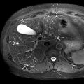
 with complete lack of internal echoes, imperceptible walls, and posterior acoustic enhancement
  . The gallbladder
  was normal.
 in addition to multiple smaller cortical cysts
  . Large cysts may produce distension, pain, or spontaneous hemorrhage.
 . Color Doppler should be used to confirm that an anechoic lesion is not vascular.
 . Acoustic enhancement
  is well seen despite the small size of the cyst.