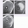
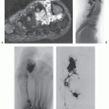
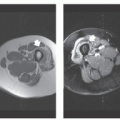
The evaluation of soft tissue tumors has undergone a dramatic change with the advent of computed tomography (CT) and magnetic resonance (MR) imaging. Despite these sophisticated techniques and the increasing number of lesions that may have a characteristic imaging appearance (e.g., lipoma, hemangioma, subacute hematoma, pigmented villonodular synovitis), many lesions remain nonspecific. Initially, investigators noted that the majority of lesions were nonspecific, with a correct histologic diagnosis reached on the basis of imaging studies alone in only approximately 25% to 35% of cases (
1,
2,
3,
4). More recently, this number has increased to more than 50% of cases with an accuracy of 85% in differentiating between benign and malignant lesions (
5).
Despite these positive developments in diagnosis, it is often not possible to establish a meaningful differential diagnosis for nonspecific soft tissue lesions or to reliably determine whether they are benign or malignant. Unlike their intraosseous counterparts, soft tissue lesions cannot be reliably assessed by evaluating their growth rate or physical parameters. In cases with a nonspecific imaging appearance, knowledge of tumor prevalence, along with the patient’s age and the lesion’s location, allows one to develop a suitably ordered differential diagnosis.
This chapter presents the results of a retrospective analysis of 31,047 soft tissue tumors seen in consultation by the Department of Soft Tissue Pathology, Armed Forces Institute of Pathology, Washington, DC, during the 10-year period starting January 1, 1980 (
6,
7). The purpose of this analysis was to (a) determine the relative prevalence, age at presentation, sex distribution, and skeletal distribution of soft tissue tumors and (b) ascertain the relative frequency of these tumors in specific anatomic locations and age groups among a population of patients in a large pathologic consultation service.
Only mesenchymal lesions originating in soft tissue were included in the study. Intra-abdominal and retroperitoneal lesions were also included when the lesions were not thought to originate in the bowel or abdominal viscera. Hence, leiomyosarcoma of the vena cava was included, whereas an angiosarcoma of the spleen was not. Lesions arising in the chest and abdominal walls and the paraspinal region were also included, as they are frequently within the purview of the musculoskeletal radiologist.
All soft tissue tumors and tumor-like lesions were placed in one of 121 major diagnostic categories. For purposes of analysis, all lesions were placed in one of 10 locations: hand and wrist, upper extremity, proximal limb girdle (axilla and shoulder), foot and ankle, lower extremity, hip and buttocks region, head and neck, trunk, retroperitoneum, and other lesions. This last category included lesions coded as abdomen, pelvis, mediastinum, or location unknown.
In total, the records of 42,490 lesions occurring in 38,484 patients were reviewed. Multiple lesions were seen in 639 patients (1.7%), including 592 patients with two lesions, 39 patients with three lesions, 7 patients with four lesions, and 1 patient with five lesions. Sequential
biopsy specimens were found in 3,311 cases. A total of 39,179 soft tissue tumors (and tumor-like masses) were available for detailed analysis. From this group, 8,132 nonmesenchymal lesions were excluded.
There were 12,370 malignant mesenchymal lesions. More than 80% were classified into seven pathologic diagnoses: malignant fibrous histiocytoma (24%), liposarcoma (14%), leiomyosarcoma (8%), malignant peripheral nerve sheath tumor (6%), dermatofibrosarcoma protuberans (6%), synovial sarcoma (5%), and fibrosarcoma (5%); 12% could not be further classified. There were 18,677 benign mesenchymal lesions. Approximately 70% of benign lesions were classified into eight pathologic diagnostic categories: lipoma and lipoma variants (16%), fibrous histiocytoma (13%), nodular fasciitis (11%), hemangioma (8%), fibromatosis (7%), neurofibroma (5%), schwannoma (5%), and giant cell tumor of tendon sheath (4%).
A summary of the malignant and benign lesions is presented in
Tables 2.1 and
2.2. A summary of the age and sex of the patients as well as the distribution of lesions for all histologic diagnoses is shown in
Tables 2.3,
2.4,
2.5,
2.6,
2.7,
2.8,
2.9,
2.10,
2.11,
2.12,
2.13 and
2.14.
The patient age and lesion location were known in 26,854 patients. For this group, the number and percentage of the seven most common malignant and benign lesions for each age and location are shown in
Tables 2.15,
2.16,
2.17,
2.18,
2.19,
2.20,
2.21,
2.22 and
2.23. All liposarcoma and fibrosarcoma subtypes have been grouped together for this analysis, as have all hemangiomas, lymphangiomas, and superficial and deep fibromatoses. Lipoma, lipomatosis, spindle cell lipoma, pleomorphic lipoma, and intramuscular lipoma have been combined and classified as lipoma. In total, 31 malignant and 52 benign diagnostic categories were used for this analysis.
The referral nature of the cases may introduce a bias for difficult case material and may be responsible for the relatively high percentage of malignancies (approximately 40%). This is greater than the 16% noted by Lattes (
8) in citing the records of Columbia University during the 45.5 years from February 1, 1906, to September 1, 1951 (1,349 malignant and 7,337 benign lesions), and considerably greater than the 5% reported by Myhre-Jensen (
9) during the 7-year period from April 1970 to April 1977 (72 malignant and 1,331 benign lesions) at the University Institute of Pathology, Aarhus, Denmark. Because of the increased number of malignancies, benign and malignant lesions have been considered separately in order to reflect accurately their relative prevalence.
In 2002, the World Health Organization (WHO) revised its classification of soft tissue tumors, incorporating new cytogenetic and molecular genetic information (
10). The WHO updated this again in 2013 (
11); however, these updates have had little effect on the tabular data. Accordingly, for this analysis we have retained the original tumor nomenclature and doubt this will cause any difficulty. That is not to say that there have not been changes in nomenclature. For example, the lesion listed in the tables as
MFH (malignant fibrous histiocytoma) now been designated
undifferentiated pleomorphic sarcoma. The major diagnostic categories, however, are not significantly changed and variations in nomenclature are explained in the written chapter text.


 Get Clinical Tree app for offline access


