Abstract
Objective
Despite aggressive therapy, approximately 50% of patients with neuroblastoma (NB) fail to respond, and survivors endure lifelong toxicities. Sonopermeation increases drug uptake via cell bilayer disruption through focused ultrasound and microbubbles (MBs)—gas-filled, sound sensitive lipid spheres. MB response to a given ultrasound pulse (cavitation) varies according to MB size. We asked whether size-sorted MBs (SSMB) 4 to 5 µm in diameter will more consistently and predictably enhance doxorubicin uptake, compared with polydisperse MBs (PMB, 0.5–10 µm in diameter), thereby increasing drug delivery to NB xenografts.
Methods
Human NB cells were implanted into the left kidney of nude mice and grown for 5 to 6 wk. Mice received sonopermeation alongside either PMB or SSMB at low (0.6 MPa) or high (2 MPa) negative pressures. Some mice also received different chemotherapy agents (doxorubicin, etoposide or cyclophosphamide). Circulating tumor cells were assessed by flow cytometry 1 h after treatment. Survival was assessed for up to 21 d, a subset of mice was euthanized 24 h after treatment for histological assessment of apoptosis, vascular lumen size and tight junctions.
Results
Tumors treated with SSMB and high pressure showed synergy with liposomal doxorubicin (L-DOX) owing to increased vascular lumen and disruption of tight junctions, resulting in drug uptake, apoptosis, lack of tumor growth and increased survival. We found no difference in the numbers of circulating tumor cells.
Conclusion
Sonopermeation with SSMB at 2 MPa synergizes with L-DOX delivery, increasing apoptosis, perfusion and vascular permeability, suggesting that SSMB sonopermeation at high pressure is promising for NB-targeted treatment, especially in combination with L-DOX.
Introduction
Neuroblastoma (NB) is the most common malignant extracranial solid tumor of childhood [ , ]. It is also the most common malignancy diagnosed within the first year of life, with a median age at diagnosis of 19 months [ ]. NB is an embryonal tumor of the sympathetic nervous system that can be found anywhere along the paraspinal sympathetic chain or in the adrenal medulla [ , ]. High-risk tumors account for approximately one-half of all NB diagnoses and require an intensive treatment regimen with multiple rounds of high-dose chemotherapy, immunotherapy, stem cell transplantation and surgery to resect or debulk the tumor [ ]. Despite the use of aggressive multimodal treatment regimens, high-risk NB has a five year disease-free survival of approximately 50% and accounts for approximately 15% of malignancy-associated pediatric deaths [ , ]. Additionally, this intense therapy often results in acute and long-term complications from toxicities owing to systemic effects [ ].
The current standard of care in NB treatment includes chemotherapy with doxorubicin, cyclophosphamide and etoposide, and other small molecule drugs, which exposes patients to acute and/or long-term toxicities. For example, doxorubicin can elicit cardiotoxicity [ ] up to 20 y after administration [ ]. Therefore, a targeted strategy allowing a decrease in systemic drug exposure while maintaining efficacy could be of great benefit to patients.
Irregular tumor vasculature poses a challenge when delivering intravenous chemotherapy to solid tumors [ , ]. We have shown that focused ultrasound (US) driving microbubble (MBs) (gas-filled, acoustically sensitive, lipid spheres) cavitation can enhance targeted delivery of high dose liposomal doxorubicin (L-DOX) [ ]. Low-intensity US will induce MB expansion and contraction (stable cavitation) whereas high-intensity US will induce violent MB collapse (inertial cavitation) [ , ]. MB cavitation has been shown to elicit membrane disruption, endocytosis and the formation of membrane pores [ , ]; this effect is referred to as “sonoporation” or “sonopermeation.”
The influence of MB size on the delivery of therapeutics has been thoroughly investigated in applications like opening the blood–brain barrier, indicating that size [ , ] and/or the overall gas volume of MBs can impact drug uptake [ ]. However, research on the effects of MB size within tumors in vivo is scarce, with no studies on NB specifically identified by our team. As research advances, understanding how MBs affect specific tumor types and their unique responses—which may differ across tumor types or as the disease progresses—becomes increasingly crucial.
Polydisperse MBs (PMBs) range from 0.5 to 10.0 µm. We have demonstrated successfully PMB sonopermeation with and without L-DOX [ ] in a NB xenograft model. Here, we compared PMBs with size-sorted MBs (SSMB) 4 to 5 µm in diameter, which have been shown to have increased efficacy when compared with PMB in examination of the blood–brain barrier [ , , ]. Our studies compare drug delivery and biological effects of SSMB with PMB of equal composition using 2.0 and 0.6 MPa peak negative US pressures in a well-characterized NB xenograft model. Finally, we tested the effects of PMBs and SSMBs on free drugs, including doxorubicin, etoposide and cyclophosphamide. We hypothesized that a more uniform distribution of larger size MBs using SSMB would generate bioeffects more favorable for enhanced drug accumulation in NB. Overall, our study highlights the potential use of SSMBs to promote L-DOX delivery in the treatment of NB to improve delivery of doxorubicin or other encapsulated drugs, which currently are not used as standard of care in NB treatment.
Materials and methods
Cell culture and NB cell lines
NGP cells (Leibniz Institute DSMZ, Braunschweig, Germany) expressing luciferase were maintained in RPMI (Gibco, Grand Island, NY, USA) with 10% fetal bovine serum (Gibco), 2.5% Geneticin (Gibco) and 5% penicillin/streptomycin (Gemini Bio Products, West Sacramento, CA, USA) in 5% CO 2 at 37°C. Cells were detached with Versene (Gibco) or Trypsin (Gibco) 0.05%. LA1-55n, LAN-5, SK-N-AS, BE2, SHEP and SH-SY5Y were kindly donated by Susan Cohn and Mark Applebaum (University of Chicago) and maintained in equal conditions, except for Geneticin.
MB development
For studies at 2 MPa , both SSMB (Supplied by AM Labs, Sudbury, MA, USA) and PMB were prepared from a lipid suspension comprised of 1, 2-distearoyl-sn-glycero-3-phosphocholine and 1,2-distearoyl-sn-glycero-3-phosphoethanolamine-N-[methoxy(polyethylene glycol)-2000] (Avanti Polar Lipids, Alabaster, AL, USA) at molar ratio of 90:10 mol% 1, 2-distearoyl-sn-glycero-3-phosphocholine to 1, 2-distearoyl-sn-glycero-3-phosphocholine and 1,2-distearoyl-sn-glycero-3-phosphoethanolamine-N-[methoxy(polyethylene glycol)-2000] in a phosphate-buffered saline (PBS) solution at pH 7.4. The constituent gas was perfluorobutane (FluoroMed, Round Rock, TX, USA).
For studies at 0.6 MPa, the formulation of the bubbles were slightly altered by the supplier. Both SSMBs (supplied from AM Labs) and PMBs were prepared from a lipid suspension comprising of 1,2-dipalmitoyl-sn-glycero-3-phosphocholine and 1,2-dipalmitoyl-sn-glycero-3-phosphate (DPPA), 1,2-dipalmitoyl-sn-glycero-3-phosphoethanolamine-N-[methoxy(polyethylene glycol)-5000] (Avanti Polar Lipids) at a molar ratio of 82:10:8 mol% 1,2-dipalmitoyl-sn-glycero-3-phosphocholine:DPPA:1,2-dipalmitoyl-sn-glycero-3-phosphate (DPPA), 1,2-dipalmitoyl-sn-glycero-3-phosphoethanolamine-N-[methoxy(polyethylene glycol)-5000] in a PBS solution containing 10 vol% propylene glycol and 10 vol% glycerol at pH 6.5. The constituent gas was octafluoropropane (FluoroMed). SSMBs were prepared using Advanced Microbubbles’ (Newark, CA, USA) proprietary MB size isolation protocol; PMB was prepared by the shaking method [ ]. Size distribution analysis of the produced MBs was measured using a Multisizer III coulter counter (MS3, Beckman Coulter, Brea, CA, USA).
NB xenografts
The NB xenograft tumor model consists of 1 × 10 6 MYC-N-amplified NGP cells implanted into the kidneys of 6- to 8-wk-old female NCr nude athymic mice. These xenografts are commonly used as a model of poor prognosis NB requiring high-dose chemotherapy. We have previously described this model in depth [ ]. This tumor model produces metastases to multiple organs relevant to NB that can be detected by IVIS (Roche, Basel, Switzerland) [ ]. Therefore, we excluded mice with detectable metastatic lesions as previously described [ ] before circulating tumor cell (CTC) studies. All experiments were approved by UT Dallas and University of Chicago IACUC. Animal numbers were estimated based on previous experimental data, anticipating an error of 0.8 and significance of 0.05, anticipating a 30% difference. For survival experiments, animals were excluded if they reached the maximum allowed tumor size before the end of the experiment or if we observed adverse effects as stated in our ACUP. Animals are always assigned to different treatment groups randomly, sorted according to starting tumor size, ensuring starting tumor sizes are equal.
Sonopermeation: Low-pressure US (0.6 MPa)
Mice were sonopermeated under isoflurane or ketamine/xylazine anesthesia. MBs were infused at a fixed concentration as previously described [ ]. A therapeutic US machine (SoundCare Plus, New York, NY, USA) with a 1 cm diameter was used to attain a maximum pressure of approximately 0.6 MPa at the tumor site (measured using a hydrophone as described by Bellary et al. [ ]). The input settings on the SoundCare plus system were 10% Duty Cycle, 1 MHz frequency and 3 W/cm 2 . US was applied during MB infusion with 10⁸/mL MB with intermittent cycles of 5 s on, 5 s off for 5 min.
High-pressure US (2 MPa)
The equipment and settings were equal to lower pressure described elsewhere in this article. To achieve a higher pressure at the tumor site, a custom lens and cone system was 3-D printed and affixed to the therapeutic US machine to attain a maximum pressure of approximately 2 MPa in the focal zone (as described by Bellary et al. [ ]).
Drugs
For studies using higher pressures, mice received 1 mg/kg L-DOX via tail vein injection during sonopermeation (n = 6 per group). This dose was based on the equivalent of human dosage, corresponding with approximately 5 mg/m 2 . Untreated controls received no treatment. For studies using low pressure, mice received doxorubicin (not liposomal) at 4 mg/kg (n = 6 per group), cyclophosphamide at 20 mg/kg (n = 3–4 per group) or etoposide at 0.3 mg/kg (n = 3 per group) immediately after or without sonopermeation (n = 3–6 per group), selected to mimic the current standard of care for these drugs.
CTC assays
NGP cells expressing luciferase and transfected with green fluorescent protein (GFP) PCCL plasmids were used for these studies only. Cells were sorted for GFP expression to ensure only transfected cells were injected into the kidneys of nude mice as described elsewhere in this article. All tumors were implanted the same day, using the same batch of cells, which were confirmed to be 86% GFP positive the day of implantation using flow cytometry. Mice were randomized for treatment using IVIS (Roche). Mice with detectable metastasis were eliminated from the study and analysis. Under ketamine/xylazine anesthesia before sonopermeation, 20 to 100 µL of blood was collected retro-orbitally into tubes containing ethylenediaminetetraacetic acid. At 1 h after sonopermeation, 300 to 500 µL of blood was harvested via cardiac puncture immediately before euthanasia, while animals were still under anesthesia. Ethylenediaminetetraacetic acid-treated blood samples were mixed 1:10 with ACK Erythrocyte Lysing Buffer (Quality Biological Inc, Gaithersburg, MD, USA) and gently shaken at 4°C for 10 min. The samples were centrifuged at 2,000 RPM at 4°C for 10 min, and the resulting pellet was resuspended in PBS. Single cell suspension was acquired using LSR-Fortessa 4-15 HTS recording 1 × 10 5 events per sample, visualized using BD FACSDiva Software and analyzed on FlowJo Software V10 (BD). After side scatter and forward scatter sorting were conducted to visualize white blood cell populations, debris and cell aggregates were gated out. GFP-positive tumor cells (CTCs) were counted using the 488 nm filter, using a single-cell suspension of the mouse’s primary tumor as an internal positive control to ensure loss of GFP had not occurred during tumor growth. CTCs were analyzed using paired t -tests (repeated measures) on GraphPad (Prism, La Jolla, CA, USA).
Tumor growth curves and survival studies
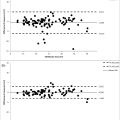
Tumor volumes were measured using calipers at the start of the experiment and every other day after treatment over 7 d using 2 Mpa US settings ( Fig 1 ). Tumor growth curves are plotted over time as a percentage of initial tumor size (approximately 1 g). All mice were euthanized when controls reached 50% growth from day 0; individual mouse curves are graphed to visualize growth ( Fig 1 B). Statistics for growth curves were performed using two-way repeated measures in GraphPad (Prism) on the groups that survived 7 d after treatment. In contrast, mice receiving 0.6 MPa US pressure ( Fig 4 a, 4 b, 4 c) were followed for up until they reached the maximum allowed tumor size (50% of day 0), thus comparing survival. Individual growth curves are not depicted owing to lack of difference between groups. Survival or Kaplan-Meier curves, contributing to survival, were analyzed on GraphPad (Prism), and significance was determined using the Log rank (Mantel Cox) test followed by a Tukey’s multiple group comparison test.

Tissue processing
Tumors were harvested 24 h after treatment and flash frozen in OTC (Vector Labs, Burlingame, CA, USA) or fixed in 4% paraformaldehyde and embedded in paraffin. We mounted 5 µm sections on slides for analysis. For safety studies in Figure 5 a, organs were harvested 1 h after sonopermeation. All other tissues analyzed by histology, enzyme-linked immunosorbent assay (ELISA) or polymerase chain reaction (described elsewhere in this article) were harvested 24 h after treatment.
Immunostaining
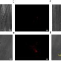
Fresh frozen tissue sections or paraffin embedded tissues were processed as described elsewhere in this article. Antibodies used were anti-zonula occludens-1 (ZO-1) (4 μg/mL; ZMD.437, Invitrogen, Carlsbad, CA, USA) with goat anti-rabbit Alexa Fluor-488 as a secondary antibody (Life Science), anti-αSMA-Cy3 (1:1,000; Sigma C6198; Sigma, St. Louis, MO, USA), and Isolectin-B4 red 568 (1:100, Invitrogen). Slides were mounted with VECTASHIELD Vibrance Antifade Mounting Medium with 4′,6-diamidino-2-phenylindole (DAPI) (Vector Laboratories). Slides were examined under an Axioskop2 microscope (Zeiss, Jena, Germany) and images analyzed in Image J (NIH, Bethesda, MD, USA), with an excitation of 561 nm and emission of 594 nm for IsolectinB4, excitation of 480 nm and emission of 535 nm for ZO1 and an excitation of 358 nm and emission 461 for DAPI. Hematoxylin and eosin staining was performed by UChicago HTRC core. Whole slide scans of 0.6 MPa organs were performed and analyzed using Pannoramic Viewer (Leica, Wetzler, Germany). Pictures of 2 MPa organs were captured at 10× magnification with a Nikon Ti2 and processed using ImageJ (NIH). Doxorubicin was localized through autofluorescence in fresh frozen slides as described [ ] without mounting using a Marianas Confocal Microscope (Zeiss) with an excitation of 561 nm and emission of 594 nm, at 20× magnification. Apoptosis was assessed with the terminal deoxynucleotidyl transferase dUTP nick end labeling (TUNEL) ApopTag kit (Millipore, Burlington, MA, USA) as elsewhere in this article [ ]. Whole slide scans (VS200, Olympus [Tokyo, Japan], with a 405 nm, 488 nm, 561 nm,= and 638 nm laser combiner, 20×) were used to scan and quantify the areas of TUNEL positivity. These areas were delineated using the freehand polymer measuring tool (area in square micrometers) from Olympus Olyvia at 20× magnification, added and divided by the total tumor tissue area analyzed using DAPI. Tumor edges where apoptosis is common in untreated tumors were excluded, as well as any remnants of normal kidney structures (not tumor tissue). Data were analyzed on Prism (GraphPad) using an ordinary one-way analysis of variance (ANOVA) followed by Tukey’s multiple comparisons test. All images were assembled in Illustrator (Adobe, San Jose, CA, USA).
Vascular lumen
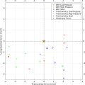
Fresh frozen sections were fixed in acetone at 4°C, blocked with CAS block (Invitrogen), and incubated with anti-endomucin (sc-65494, 1:100; Santa Cruz Biotechnology, Dallas, TX, USA). Anti-rat (BA-9400, Vector Labs) secondary was then followed by streptavidin-horseradish peroxidase (Vector Labs) and AEC substrate (sk-4205 Vector Labs). Last, slides were counterstained using hematoxylin (Vector Labs). Slides were scanned and lumens measured in Panoramic viewer (Leica) using the distance tool. The widest dimension within each endomucin positive blood vessel was selected and at least 30 measurements were taken per tissue (n = 3 per group). Two-way ANOVA followed by Tukey’s comparison was performed in Prism (GraphPad).
Protein sample preparation
Tumor samples were homogenized with a Polytron PT1200E in RIPA lysis buffer (Sigma-Aldrich) containing 1 mM PMSF (Pierce, Appleton, WI, USA) and Cocktail Protease Inhibitor Tab (Roche), at 4°C for 20 min. After centrifugation (14,000 RPM for 20 min at 4°C), protein concentrations were measured with a bicinchoninic acid assay (Thermo Fisher Scientific, Waltham, MA, USA) following the manufacturer’s instructions.
ELISA for ZO-1
High binding (Costar, Washington, DC, USA) plates were coated with 2 μg/mL anti ZO-1 ZMD.437 (Invitrogen) polyclonal antibody diluted in a coating buffer (Thermo Fisher Scientific) and incubated overnight. After blocking (Thermo Fisher Scientific), 7.9 mg/μL protein of tumor homogenates or controls were incubated for 1 h. After washing, the detection antibody solution (0.5 μg/mL anti-ZO-1 ZO-1.1412, Invitrogen) was incubated for 1 h in blocking buffer, followed by 1 h of streptavidin-horseradish peroxidase (Thermo Fisher Scientific) and TMB substrate (Thermo Fisher Scientific) for colorimetric detection. The reaction was stopped after 30 min, and absorbance was quantified using the SpectraMaxi3x at 450 nm. NGP cells were used as a positive control, and RIPA buffer served as a negative control. For ELISA, a two-tailed Student’s t -test was performed after confirming normality of data using Prism (GraphPad).
Sodium dodecyl sulfate–polyacrylamide gel electrophoresis
We loaded 25 mg of denatured proteins in an 8% gel using a Spectra protein ladder (Sigma) using a Bio-Rad mini chamber. The proteins were transferred to a nitrocellulose membrane (Li-Cor, Lincoln, NE, USA), followed by anti-ZO-1 (ZMD.437, Invitrogen) or β-actin (Cell Signaling 8H10D10 1:1,000 dilution) as loading controls. Secondary antibodies used were goat anti-rabbit 800CW and goat anti-mouse 680CW (1:1,000, IRdye, Li-COR). IRDyes were captured using Azure Biosystems. The ratio of ZO-1 to β-actin was calculated with ImageJ (NIH). There are no statistics because the goal was to confirm the presence or absence of ZO-1.
Animal groups
Two tables detail all animal groups with treatment, timepoints, independent and dependent variables. Table 1 details animals under 2 MPa US pressures, and Table 2 details animals receiving 0.6 MPa US pressures.
| Group | MB Type | Drug | End point |
|---|---|---|---|
| #1 | None | None | 7 d |
| #2 | None | L-DOX (1 mg/kg) | 7 d |
| #3 | SSMBs | None | 7 d |
| #4 | SSMBs | L-DOX (1 mg/kg) | 7 d |
| #5 | PMBs | L-DOX (1 m/kg) | 7 d |
| #6 | none | None | 24 h |
| #7 | None | FUS only | 24 h |
| #8 | None | L-Dox (1 mg/kg) | 24 h |
| #9 | SSMBs | L-DOX (1 mg/kg) | 24 h |
| #10 | PMBs | L-DOX (1 mg/kg) | 24 h |
| #11 | SSMBs | None | 24 h |
| #12 | PMBs | None | 24 h |
Related posts:
 A Narrative Review of Image Processing Techniques Related to Prostate Ultrasound
 Assessing Intensive Care Unit Acquired Weakness: An Observational Study Using Quantitative Ultrasound Shear Wave Elastography of the Rectus Femoris and Vastus Intermedius
 Transcranial MRI-guided Histotripsy Targeting Using MR-thermometry and MR-ARFI
 Ischemia/Reperfusion Injury Enhances Accumulation of Perfluoropropane Droplets
 Application of B+M-Mode Ultrasound in Evaluating Dysphagia in Elderly Stroke Patients
 Automatic Segmentation of Quadriceps Femoris Cross-Sectional Area in Ultrasound Images: Development and Validation of Convolutional Neural Networks in People With Anterior Cruciate Ligament Injury and Surgery
Stay updated, free articles. Join our Telegram channel

Full access? Get Clinical Tree


