Abstract
This case report describes a 24-year-old male with a spinal ganglion cyst originating from the posterior longitudinal ligament (PLL) of the lumbar spine, an uncommon site for ganglion cysts. The patient exhibited symptoms of neurogenic claudication (NC), initially thought to be spinal stenosis, however MRI showed a cystic lesion instead, compressing L2-L3 nerve roots. The case illustrates the importance of differential diagnoses in spinal cystic lesions and highlights surgical excision as an effective treatment for symptomatic relief.
Introduction
Ganglion cysts commonly occur in the tendons of the wrist (60%-70%) and hand (7%-12%), but can occassionally develop in the spinal perineural connective tissue, leading to nerve root compression and associated symptoms [ ].
Differentiating ganglion cysts from other similar lesions can be challenging, and typically requires a detailed histopathologic examination due to their resemblance on imaging.
Treatment options range from pain management and physical therapy to surgical removal for neural decompression and recurrence prevention [ ].
This report describes a case of severe neurogenic claudication (NC) caused by a rare spinal ganglion cyst (SGC) originating from the PLL of the lumbar spine, and discusses its clinical and radiologic findings.
Case presentation
A 24-year-old male farm worker presented with progressively worsening lower back pain, accompanied by intermittent painful cramping and leg weakness over 6 months. Conservative medical treatment provided no relief, and his symptoms increasingly interfered with his work. He denied any preceding trauma or acute injury, reporting good health prior to the onset of symptoms.
Physical examination revealed characteristic symptoms of lumbar spinal stenosis, including low back pain, paresthesia or cramping in one or both legs, worsened with walking and improved with sitting [ ].
This postural effect, which exacerbates symptoms on spinal extension while relieving them on flexion, strongly suggests neurogenic claudication. The underlying mechanism likely involves posture-related compression, leading to neural and microvascular compromise of the cauda equina and lumbosacral nerve roots [ ].
Although surgery is not the first-line treatment for NC secondary to lumbar spinal stenosis, it is recommended for patients with severe, persistent pain or progressing neurological deficits.
Following the clinical findings, MRI with contrast (T1, T2, STIR, and DWI sequences) in sagittal and axial views was performed following gadolinium administration.
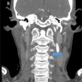
The lesion was located at L2-L3 on the anterior wall of the spinal canal in the bilateral median-paramedian region, causing significant spinal canal stenosis (narrowed to 5 mm at the affected level).
This description of the lesion should come after the next line which talks about the T1 a nd T2 weighted images, and it should read: Two internal septa created mild lobulation, with maximum lesion dimensions of 21 × 7 × 6 mm (LxWxD).The lesion exhibited or demonstrated, pick onea hypointense signal on T1-weighted images and hyperintense signal on T2-weighted images, with rim enhancement following administration of gadolinium, as shown in Fig. 1 below [ ].

Importantly, the cyst lacked synovial lining or communication with the joint cavities, excluding the possibility of a synovial cyst [ , ]. Sagittal images further demonstrated the delamination of the PLL, supporting the primary diagnostic hypothesis of a spinal ganglion cyst.
Given the patient’s persistent symptoms and failure of conservative treatment, surgical excision was performed with subsequent histologic characterization of the lesion.
There were no peri‑operative complications, and the patient reported complete symptom resolution, including the absence of low back pain and neurogenic claudication. Follow-up MRI confirmed the resolution of stenosis and restoration of normal canal width ( Figs. 2 and 3 ).

Related posts:
 A rare and life-threatening case of spontaneous hemopneumothorax presenting with severe hemodynamic instability
 A very rare case of ileocolic and appendiceal intussusception with acute appendicitis
 Hepatic and extra-hepatic hydatid cysts: A case series of radiological and clinical insights
 Mid-term follow-up of a fractured flow diverter in the internal carotid artery
 An atypical multiple autoimmune syndrome: A case report including myocarditis
 Persistence of the hypoglossal artery: A rare case report
Stay updated, free articles. Join our Telegram channel

Full access? Get Clinical Tree


