Abstract
Deep venous thrombosis can be observed in pediatric patients, especially in teenagers. There is a paucity of published treatment recommendations in this patient population. This technical note illustrates the successful utilization of aspiration thrombectomy to manage a lower extremity deep venous thrombosis in a 17-year-old boy. This shows that thrombectomy devices utilized in adult practice can be used in pediatric patients, with careful consideration of vessel size and patient specific factors.
Introduction
Deep vein thrombosis (DVT) occurs rarely in the pediatric population, at an annual rate of 10-14 per 10,000 children admitted to hospital [ ], yet incidence has been shown to be rapidly increasing [ ]. Despite its rarity, DVT carries a risk of serious complications including pulmonary embolism and post-thrombotic syndrome. Indeed, pediatric mortality secondary to DVT has been reported at 2% [ ]. There are a variety of therapeutic options for lower extremity DVT including anticoagulation, thrombolytics, and endovascular thrombectomy [ ], however these approaches are mainly described in the adult population [ ]. As such, it is challenging to apply these approaches in children due to the lack of evidence, especially with new devices. This case report describes the successful utilization of a 12 French aspiration thrombectomy device in a teenager.
Case presentation
Informed consent was obtained from the patient’s legal guardian for publication. This case describes a 17-year-old boy, previously diagnosed with T-Cell Acute lymphoblastic leukemia (ALL), who presented to the emergency department with a 1-day history of left leg swelling and fever. Upon assessment, the patient was noted to have swelling of the left lower limb, from foot to mid-thigh, with associated pain and inability to ambulate. Pedal pulses were intact, and no skin changes were noted. The patient denied shortness of breath, cough, or hemoptysis. Due to the presence of fever, blood cultures were obtained, and the patient was started on antibiotics. The patient had a recent 5-day admission due to mucositis and poor oral intake in the context of delayed intensification treatment as per specific ALL protocol. On day of discharge, the patient was noted to have predominately left-sided mild lower limb edema. This was attributed to low albumin levels given the lack of additional symptoms. The subsequent day postdischarge, the patient’s edema continued to progress, and the patient was re-admitted.
Grey scale and color-Doppler ultrasound of the left lower extremity was performed showing lack of flow from the left popliteal vein to the left common iliac vein (CIV), compatible with extensive DVT ( Fig. 1 A). The inferior vena cava (IVC) appeared patent with no signs of intraluminal thrombus. The patient was admitted, and a therapeutic dose unfractionated heparin infusion was initiated.

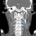
Computed Tomography of the abdomen and pelvis was performed to delineate the extension of the thrombosis. It confirmed patency of the IVC and characterized the occlusive thrombus as extending from the left common iliac vein origin to the popliteal vein ( Fig. 1 B), with marked subcutaneous and cutaneous inflammatory changes of the left upper leg. A short segment nonocclusive thrombus was additionally seen within the mid right common iliac vein. Notably, compression of the left common iliac vein was observed between the right common iliac artery and underlying lumbar vertebrae, raising the possibility of May-Thurner syndrome. Due to the extension of the thrombosis and the patient’s inability to ambulate, a multidisciplinary decision between Interventional Radiology, Oncology and the Thrombosis team was made to proceed with mechanical aspiration thrombectomy and thrombolysis of the left lower limb. It was decided not to intervene on the right leg given the small size of the thrombus.
At the time of the procedure the 12 French Penumbra Lightning TM 12 Indigo® system (Penumbra Inc, Alameda, CA, USA) was recently approved in the local practice, therefore it was decided to attempt to use this new size of catheter for the first time, considering that the size of the patient’s veins appeared to be sufficient to accommodate a 12 French vascular sheath. The procedure was performed under general anesthesia, using sterile technique, and in the prone position. The preprocedural mapping ultrasound of the left popliteal fossa showed a patent inferior portion of the popliteal vein, sufficient for access with a 12 French sheath. Under ultrasound guidance, the left popliteal vein was accessed using a 21-gauge micro-puncture needle. The access was subsequently upsized to a 12 French vascular sheath and secured using a 2-0 Prolene stitch. Through the sheath, a 0.035″ hydrophilic wire was advanced to the level of the IVC, followed by an angled Bernstein catheter over the wire. A pullback venography demonstrated diffuse clot burden throughout the left iliofemoral system and further evidence of May-Thurner syndrome ( Fig. 2 A). Venous collaterals were noted in the pelvis and thigh. After placement of a 20-cm infusion length Cragg-McNamara catheter TM (ev3, Micro Therapeutics, Inc; Irvine, CA, USA), pulse-spray tPA (20mg alteplase in 50 mL solution) was infused across the length of the clot burden and left in situ for 30 minutes.

Related posts:
 A rare and life-threatening case of spontaneous hemopneumothorax presenting with severe hemodynamic instability
 A very rare case of ileocolic and appendiceal intussusception with acute appendicitis
 Hepatic and extra-hepatic hydatid cysts: A case series of radiological and clinical insights
 Mid-term follow-up of a fractured flow diverter in the internal carotid artery
 An atypical multiple autoimmune syndrome: A case report including myocarditis
 Persistence of the hypoglossal artery: A rare case report
Stay updated, free articles. Join our Telegram channel

Full access? Get Clinical Tree


