Anterior Abdominal Wall
FASCIAL LAYERS
There are two layers of superficial fascia on the abdominal wall; a loose areolar fatty layer named Camper’s fascia, and deep to this a fibrous layer named Scarpa’s fascia. Camper’s fascia extends inferiorly past the inguinal ligament as the subcutaneous fat of the thigh. It extends medially past the pubic symphysis, combining with Scarpa’s fascia to form the Dartos fascia of the scrotum in males and the fatty tissue of the mons pubis and labia majora in females. Scarpa’s fascia extends deep to the inguinal ligament and posteriorly into the perineum to fuse with Colles’ fascia. It does not extend into the thigh but fuses with the fascia lata. A condensation continues onto the dorsum of the penis as a suspensory ligament.
MUSCLE LAYERS ( Fig. 5.1 )
The wall consists of three principal muscular layers. Each consists of lateral muscular fibres with aponeuroses fibres centrally. The fibrous aponeurotic components ensheath the rectus abdominis around the anterior midline.
The external oblique arises on the anterior surface of the lower eight ribs and is inserted into the linea alba, pubic crest, pubic tubercle and anterior half of the iliac crest. Between the anterior superior iliac spine and pubic tubercle the aponeurotic free border of this muscle forms the inguinal ligament. The muscle fibres run inferiorly and medially.
The internal oblique arises from the lumbar fascia, the anterior iliac crest and the lateral two-thirds of the inguinal ligament and is inserted into the costal margin, aponeurosis of the rectus sheath and the pubic crest via the conjoint tendon. Muscle fibres run superomedially at right angles to the external oblique.
The transversus abdominis arises from the deep aspect of the lower six ribs (interdigitating with the diaphragm), the lumbar fascia, the anterior iliac crest and the lateral inguinal ligament, and is inserted into the linea alba and the pubic crest via the conjoint tendon.
The rectus abdominis muscle originates from the fifth, sixth and seventh costal cartilages and inserts into the pubic crest. It has three to four tendinous intersections at intervals along its length, which adhere to the anterior rectus sheath.
The rectus sheath is formed by the aponeuroses of the external oblique, internal oblique and transversus abdominus muscles. The aponeurosis of the transversus abdominus runs deep to the rectus sheath. The aponeurosis of transversalis splits to enclose the rectus abdominus muscle. The external oblique aponeurosis is anterior to the rectus muscle. The rectus sheath fuses at the midline to form the linea alba which runs from the xiphisternum to the pubic symphysis.
The posterior lamina of the sheath, formed by the internal oblique aponeurosis (covering the posterior aspect of rectus muscles), terminates inferiorly halfway between the umbilicus and pubis symphysis, whereas the anterior lamina covers the rectus to its insertion into the pubic bone. The free end of the posterior lamina of the sheath is the arcuate line of Douglas or linea semilunaris . Superior to the arcuate line, the posterior rectus sheath is made up of the aponeurosis of transverses abdominis and some of the aponeurosis of internal oblique; below the arcuate line there is only transversalis fascia. The arcuate line marks the level at which the inferior epigastric vessels enter the rectus sheath.
A layer of fat separates the muscles of the anterior abdominal wall from the peritoneum and the abdominal contents. This is referred to as extraperitoneal or properitoneal fat.
UMBILICAL LIGAMENTS
The median umbilical ligament is an unpaired midline structure. It runs from the bladder fundus, along the deep aspect of the anterior abdominal wall to the umbilicus. It consists of the fibrous remnant of the obliterated fetal urachus. It is covered by a fold of peritoneum called the median umbilical fold.
The medial umbilical ligaments are the paired fibrous remnants of the obliterated umbilical arteries and run from the internal iliac artery to the umbilicus on the deep surface of the anterior abdominal wall. These are also covered by folds of peritoneum called the medial umbilical folds.
The lateral umbilical fold refers to the fold of parietal peritoneum that covers the inferior epigastric artery as it passes superomedially from the external iliac artery to enter the rectus sheath.
RADIOLOGICAL FEATURES OF THE ANTERIOR ABDOMINAL WALL
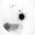
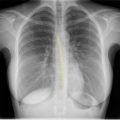
Plain Films of the Abdomen ( Fig. 5.1 )
The muscle layers of the anterior abdominal wall are outlined, especially in obese individuals, between the subcutaneous fat line and the properitoneal fat line. Clearly seen fat lines indicate a lack of oedema in these areas; in 18% of normal radiographs the properitoneal line is absent.

- 1.
Abdominal wall musculature
- 2.
Properitoneal fat line (extraperitoneal fat layer)
- 3.
Inferior border of the right lobe of the liver
- 4.
Gas in the hepatic flexure
- 5.
Right twelfth rib
- 6.
Splenic flexure
- 7.
Left kidney
- 8.
Left psoas muscle
- 9.
Left spinous process of L5
- 10.
Spinous process of L5
- 11.
Small bowel loops
Computed Tomography (CT) of the Abdominal Wall ( Figs. 5.2 – 5.5 )
The muscle layers of the anterior abdominal wall can be seen in cross-section. Three muscles can be seen anterolaterally: the external oblique is outermost, then the internal oblique, with transversus abdominus deepest. The rectus muscle and its rectus sheath can be seen in the anterior paramedian position superficial to the other muscles.
- •
Free intraperitoneal gas may outline the umbilical ligaments and falciform ligament making them visible, thus making a diagnosis of pneumoperitoneum possible on a supine radiograph.
- •
Rupture of the bulbous urethra results in tracking of urine into the scrotum, perineum and abdominal wall deep to Scarpa’s fascia. It cannot track into the thigh due to the attachment of Scarpa’s fascia to the deep fascia of the thigh.
- •
Spigelian hernias emerge lateral to the rectus sheath at the level of the arcuate line, a relative weak point below the lower limit of the internal oblique fascia.
- •
Skin punctures lateral to the rectus muscles, or in the midline, will avoid the epigastric vessels. Catheter placement that avoids the recti is also better tolerated.


- 1.
Right lobe of the liver
- 2.
Confluence of the splenic and superior mesenteric veins to form the portal vein
- 3.
Splenic vein
- 4.
Head of the pancreas
- 5.
Second part of the duodenum
- 6.
Loops of the small bowel
- 7.
Hepatic flexure
- 8.
Descending colon
- 9.
Spleen
- 10.
Aorta
- 11.
Superior mesenteric artery
- 12.
Inferior vena cava
- 13.
Left adrenal gland
- 14.
Right kidney
- 15.
Renal cortex
- 16.
Renal pyramid in the renal medulla
- 17.
Rectus abdominis muscle
- 18.
Transversus abdominis muscle
- 19.
Latissimus dorsi muscle
- 20.
Erector spinae muscle

- 1.
Right lobe of the liver
- 2.
Aorta
- 3.
Inferior vena cava
- 4.
Right renal vein
- 5.
Left renal vein
- 6.
Left renal artery
- 7.
Superior mesenteric artery
- 8.
Third part of the duodenum passing between the aorta and superior mesenteric artery
- 9.
Loops of small bowel
- 10.
Ascending colon
- 11.
Transverse colon
- 12.
Descending colon
- 13.
Psoas muscle
- 14.
Lower end crus of the right hemidiaphragm
- 15.
Lower end crus of the left hemidiaphragm
- 16.
Right renal pelvis
- 17.
Rectus abdominis muscle
- 18.
Transversus abdominis muscle
- 19.
Internal oblique muscle
- 20.
External oblique muscle
- 21.
Latissimus dorsi muscle
- 22.
Erector spinae muscle
- 23.
Gerota’s fascia
- 24.
Fascia of Zuckerkandl
- 25.
Lateral conal fascia (fusion of Gerota’s fascia and fascia of Zuckerkandl)
The Stomach ( Figs. 5.6, 5.7 )
The stomach is J-shaped but varies in size and shape with the volume of its contents, with erect or supine position, and with inspiration and expiration.


The stomach consists of two surfaces (anterior and posterior) and two curvatures (the greater and the lesser). The lesser curve is punctuated by a notch known as the incisura.
The stomach has two orifices – the cardia and the pylorus. The cardiac orifice or cardia is so-named because of its proximity to the heart and marks the anatomical junction between the oesophagus and stomach. In clinical practice the junction is known as the gastro-oesophageal junction, oesophagogastric junction and lower oesophageal sphincter.
The dome-like projection of the stomach above the cardia is the fundus .
Between the cardia and the incisura is the body of the stomach, and distal to the incisura is the gastric antrum which leads to the pylorus . The lumen of the pylorus is referred to as the pyloric canal .
The stomach is lined by mucosa, which has tiny nodular elevations called the areae gastricae and is thrown into folds called rugae . Longitudinal folds parallel to the lesser curve are called the ‘ magenstrasse ’ meaning ‘street of the stomach’. Rugae elsewhere in the stomach are random and patternless.
There are three muscle layers in the wall of the stomach: (1) an outer longitudinal, (2) an inner circular and (3) an incomplete, innermost oblique layer. The circular layer is thickened at the pylorus as a sphincter, but not at the oesophagogastric junction. Fibres of the oblique layer loop around the notch between the oesophagus and the fundus, and help to prevent reflux.
Peritoneum covers the anterior and posterior surfaces of the stomach and is continued between the lesser curve and the liver as the lesser omentum, and beyond the greater curve as the greater omentum.
The oblique fibres are responsible for the ‘ magenstrasse ’ and can reduce the volume of the stomach, allowing fluids and food to pass directly and rapidly from the oesophagus along the lesser curve to the duodenum without filling the main lumen of the stomach. This can be seen when liquid contrast is swallowed.
ANTERIOR RELATIONS OF THE STOMACH
The upper part of the stomach is covered by the left lobe of the liver on its right and by the left diaphragm on its left. The fundus occupies the concavity of the left dome of the diaphragm. The remainder of the stomach is covered by the anterior abdominal wall.
POSTERIOR RELATIONS OF THE STOMACH
Posterior to the stomach lies the lesser sac (refer to section on peritoneal spaces for further detail). The structures that lie behind this are referred to as the stomach bed. The pancreas lies across the midportion of the stomach bed with the splenic artery partly above and partly behind it, and the spleen at its tail. Above the pancreas lie the aorta, coeliac trunk, the coeliac plexus and nodes, the diaphragm, left kidney and left adrenal gland. Attached to the anterior pancreas is the transverse mesocolon which forms the inferior part of the stomach bed.
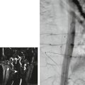
ARTERIAL SUPPLY OF THE STOMACH ( Fig. 5.8 , Figs. 5.9, 5.10 )
Branches of the coeliac trunk reach the stomach along its greater and lesser curves as follows:
- ■
The lesser curve is supplied by the left gastric artery (a branch of the coeliac trunk) and the right gastric artery (a branch of the hepatic artery).
- ■
The greater curve is supplied by the right gastroepiploic artery (from the gastroduodenal branch of the hepatic artery) and the left gastroepiploic artery (from the splenic artery).
- ■
The proximal aspect of the greater curve and the fundus are supplied by the short gastric branches of the splenic artery.


- 1.
Catheter in the aorta
- 2.
Catheter tip in the coeliac trunk
- 3.
Splenic artery
- 4.
Left gastric artery
- 5.
Common hepatic artery
- 6.
Hepatic artery proper
- 7.
Left hepatic artery
- 8.
Right hepatic artery
- 9.
Right gastric artery
- 10.
Cystic artery
- 11.
Gastroduodenal artery
- 12.
Right gastroepiploic artery
- 13.
Left gastroepiploic artery
- 14.
Posterior superior pancreaticoduodenal artery
- 15.
Anterior superior pancreaticoduodenal artery
- 16.
Dorsal pancreatic artery
- 17.
Transverse pancreatic artery
- 18.
Gastric branches of the gastroepiploic artery
- 19.
Phrenic branch of the left hepatic artery
- 20.
Contrast in right renal pelvis
- 21.
Left ureter

The arteries of the stomach anastomose freely within the stomach wall, unlike the arteries of the small and large intestine, which are end arteries.
VENOUS DRAINAGE OF THE STOMACH ( Fig. 5.11 )
Venous drainage of the stomach follows a similar pattern to arterial supply:
- ■
The right and left gastric veins drain directly into the portal vein
- ■
The short gastric and left gastroepiploic veins drain to the splenic vein
- ■
The right gastroepiploic vein drains to the superior mesenteric vein

LYMPHATIC DRAINAGE OF THE STOMACH
Lymphatic drainage follows the arterial pattern, draining to coeliac nodes which drain into the cisterna chyli:
- ■
The left gastric artery lymphatics drain directly to coeliac nodes
- ■
The right gastric artery lymphatics drain via retroduodenal nodes to coeliac nodes
- ■
The short gastric and left gastroepiploic artery lymphatics drain via nodes in the splenic hilum and behind the pancreas to coeliac nodes
- ■
The right gastroepiploic artery lymphatics drain via retroduodenal nodes to coeliac nodes
Retrograde spread of carcinoma may occur into the nodes at the porta hepatis. The extensive and complex lymphatic drainage of the stomach poses a challenge in the management of gastric cancer.
RADIOLOGICAL FEATURES OF THE STOMACH
Plain Film of the Abdomen (see Fig. 5.2 )
Erect radiographs demonstrate a wide fluid level at the gastric fundus. This is because the antrum and body contract when empty and a relatively small amount of fluid and gas in the fundus can produce a long fluid level. When the stomach is distended, two fluid levels may be seen, one in the body of the stomach and one in the antrum.
Double-Contrast Barium Meal Examination ( Fig. 5.12 )
Features of the stomach, such as the greater and lesser curves, the incisura, the cardia, fundus, body, antrum and pylorus, can be seen. The change in position of these with posture and respiration can be appreciated on fluoroscopy.

- 1.
Lesser curve of the stomach
- 2.
Greater curve of the stomach
- 3.
Longitudinal mucosal folds (‘magenstrasse’)
- 4.
Gastric rugae
- 5.
Areae gastricae
- 6.
Incisura
- 7.
Duodenal bulb (partially gas distended)
- 8.
Mucosal pattern of the nondistended duodenum
CT and Magnetic Resonance Imaging (MRI) of the Stomach ( Figs. 5.13, 5.14 )
The thickness of the stomach wall varies considerably depending on the degree of distension and can appear thickened in the fasting state.

- 1.
Left lobe of the liver
- 2.
Right lobe of the liver
- 3.
Right hepatic vein
- 4.
Fissure for the ligamentum venosum
- 5.
Contrast medium in the stomach
- 6.
Spleen
- 7.
Descending aorta
- 8.
Azygos vein
- 9.
Hemiazygos vein
- 10.
Splenic flexure
- 11.
Left lung
- 12.
Left diaphragm
- 13.
Rectus abdominis muscle
- 14.
External oblique muscle
- 15.
Serratus anterior muscle
- 16.
Latissimus dorsi muscle
- 17.
Erector spinae muscle
- 18.
Oesophagogastric junction

- 1.
Left lobe of the liver: lateral segment
- 2.
Left lobe of the liver: medial segment
- 3.
Right lobe of the liver
- 4.
Caudate lobe of the liver
- 5.
Fissure for ligamentum teres
- 6.
Interlobar fissure (for ligamentum venosum)
- 7.
Inferior vena cava
- 8.
Portal vein
- 9.
Right branch of the portal vein
- 10.
Gallbladder
- 11.
Hepatic artery
- 12.
Abdominal aorta
- 13.
Splenic artery
- 14.
Spleen
- 15.
Splenic vein
- 16.
Left crus of the diaphragm
- 17.
Left adrenal gland
- 18.
Right adrenal gland
- 19.
Stomach
- 20.
Splenic flexure
- 21.
Linea alba (aponeurosis of the rectus sheath)
The mucosa of the stomach enhances with intravenous contrast and the stomach layers are best appreciated in the arterial phase of contrast enhancement, This enhancement is best seen when there is no positive contrast in the stomach.
Ultrasound Studies of the Stomach
Ultrasound is limited by the presence of intraluminal gas in the stomach. Structures in the stomach bed cannot be seen through the stomach if this is filled with gas. If the stomach is gas free or fluid filled, then the pancreas and the aorta and coeliac trunk can be seen well.
- •
In the infant, ultrasound is used in the study of the pylorus in cases of suspected pyloric stenosis. Pyloric muscle thicknesses greater than 3 mm and muscle lengths greater than 14 mm are considered abnormal.
- •
The stomach can be filled with gas using effervescent granules to eliminate confusing rugal patterns, and to push other bowel loops out of the field of interest.
- •
The fundus of the stomach may project posteriorly if redundant and may mimic an adrenal mass on CT and MRI.
- •
Food in the stomach can appear as a pseudomass, especially in the dependent portion.
The Duodenum ( Figs. 5.15, 5.16 )
The duodenum extends from the pylorus, curving in a C shape around the head of the pancreas to end at the duodenojejunal flexure, where transition to the small bowel proper is marked by the assumption of a longer, more mobile mesentery.


- 1.
Sacculation
- 2.
Collapsed transverse colon
- 3.
Duodenal bulb
- 4.
Stomach
- 5.
Splenic flexure
- 6.
Duodenojejunal junction
- 7.
Jejunum
- 8.
Ileum
The first 2.5 cm of duodenum, like the stomach, is attached to the greater and lesser omentum. The remainder of the duodenum is retroperitoneal, with a short mesentery and, as a result, it is less mobile. Its anterior surface is covered by peritoneum, except where the second part is crossed by the transverse mesocolon and where the third part is crossed by the superior mesenteric vessels in the root of the mesentery.
It is described as having four parts: the first (or superior), second (or descending), third (or horizontal) and fourth (or ascending). These measure approximately 2 cm, 8 cm, 8 cm and 4 cm, respectively. The first part is at the level of L1 lumbar vertebra, the second at L2, the third at L3, and the fourth ascends again to L2 level.
- ■
The first part (superior) , called the duodenal bulb/cap, passes posterosuperiorly over a distance of approximately 2 cm on the transpyloric plane at L1. It is covered by the peritoneum, with the lesser omentum extending from its superior edge and the greater omentum from its inferior. The common bile duct, the portal vein and the gastroduodenal artery pass behind the first part of the duodenum and separate it from the inferior vena cava (IVC). Inferiorly it is in contact with the pancreatic head.
- ■
The second part (descending) of the duodenum is approximately 8 cm long. It runs inferiorly around the head of the pancreas at the level of the L2 and L3 vertebral bodies. The common bile duct (CBD) and the main pancreatic duct open into posteromedial wall of the second part of the duodenum at the ampulla of Vater/dueodenal papilla. This is guarded by the sphincter of Oddi. An accessory pancreatic duct (of Santorini), if present, opens proximal to this.
- ■
This part of the duodenum is crossed by the transverse mesocolon anteriorly. As a result, its upper half is supracolic and has the liver as an anterior relation. Its lower half is infracolic and has loops of jejunum anteriorly. Its posterior relations are the right kidney and it is in contact with the pancreatic head medially.
- ■
The third part (horizontal) of the duodenum runs for approximately 8 cm horizontally across the anterior aspect of the L3 vertebra, the IVC and aorta. Its posterior relations include the right psoas muscle, ureter and gonadal vessels of the posterior abdominal wall. Anteriorly it is crossed by the root of the mesentery and the superior mesenteric vessels. The head of the pancreas is in contact with its superior border.
- ■
The fourth part (ascending) of the duodenum passes upwards before turning abruptly anteriorly. Its superior turn is suspended by a peritoneal fold which descends from the right crus of the diaphragm, called the ligament of Treitz. At this ligament the small bowel assumes a longer mesentery, marking the transition to the jejunum. The ligament is not easily identified radiologically, its lower limit is inferred by the duodenojejunal flexure, normally the highest point of the duodenum. The DJ flexure normally lies on the left side of the aorta, superficial to the left psoas muscle and posterior to the stomach. The inferior mesenteric vessels raise another peritoneal fold lateral to the fourth part of the duodenum.
ARTERIAL SUPPLY (see Fig. 5.15 )
The first part of the duodenum is supplied by the right gastric and the right gastroepiploic arteries.
The superior pancreaticoduodenal artery , a branch of the gastroduodenal artery from the hepatic, supplies the first half of the second part.
The remainder of the duodenum is supplied by the inferior pancreaticoduodenal artery , the first branch of the superior mesenteric artery (SMA). At the midpoint of the second part of the duodenum, therefore, there is a transition from supply by the coeliac trunk to supply by the SMA, representing a transition from foregut to midgut.
VENOUS DRAINAGE OF THE DUODENUM
The first part of the duodenum drains to the prepyloric vein (of Mayo), which lies on the anterior surface of the pylorus, and thence to the portal vein. The remainder drains to veins that correspond to the arteries. These drain to the portal and superior mesenteric veins.
LYMPHATIC DRAINAGE OF THE DUODENUM
Pancreaticoduodenal nodes drain to pyloric nodes and to coeliac nodes.
RADIOLOGICAL FEATURES OF THE DUODENUM
Barium Studies of the Duodenum ( Figs. 5.12, 5.16 )
The duodenum is usually examined radiologically as part of a double-contrast barium meal examination (see Fig. 5.12 ).
The first part of the duodenum passes posteriorly as well as superiorly, therefore it is best evaluated with the right side raised in a right anterior oblique view (it is foreshortened in anteroposterior, [AP] views).
The duodenal bulb may be indented by the normal gallbladder. The bulb has thin mucosal folds that are parallel, or parallel in spiral, from base to apex. Circular valvulae conniventes begin in the second part of the duodenum.
The ampulla is visualized in two-thirds of normal examinations and an opening of an accessory pancreatic duct in less than one-quarter. The accessory duct opens more anteriorly than the main duct.
The third part of the duodenum is indented by the aorta posteriorly and superior mesenteric vessels anteriorly.
- •
A complete upper gastrointestinal barium examination should extend to the ligament of Treitz.
- •
Normally the ligament of Treitz marks the highest part of the fourth part of the duodenum and marks the transition to jejunum. The DJ flexure should lie to the left of the first part of the duodenum. It should be at the same height or above the duodenal bulb and lie in a posterior, retroperitoneal location. An abnormal position of the ligament of Treitz/DJ flexure indicates intestinal malrotation. In such cases, jejenum and small bowel lie predominantly on the right, and large bowel lies predominantly to the left, of midline.
Ultrasound
Gas in the duodenum may hinder visualization of other organs, particularly the common bile duct, which runs behind the first part. The duodenum may be identified adjacent to the head of the pancreas by ultrasound.
The Small Intestine
The small intestine begins at the duodenojejunal flexure and ends at the ileocaecal junction. It has a variable length, averaging 6 m. The mesentery in continuity with the small bowel extends from the left side of L2 to the right sacroiliac (SI) joint ( Fig. 5.17 ). The small intestine is very mobile and lies in mobile coils in the central abdomen. The proximal two-fifths of the small intestine is called the jejunum and the distal three-fifths the ileum, although the boundary between these is not well defined.

Circular mucosal folds – known as valvulae conniventes or plicae semilunaris – are seen in the duodenum and continued in the small intestine, although they are less prominent distally and may be absent in distended views on barium studies.
Lymphoid follicles found in the mucous membrane throughout the intestinal tract become increasingly more numerous along the length of the small intestine. In the distal ileum they become aggregated together into patches called Peyer’s patches . They are oval in shape and found on the antimesenteric border of the ileum.
Differences between the jejunum and ileum are outlined in Table 5.1 .
| Jejunum | Ileum | |
|---|---|---|
| Diameter | Wider (3.0–3.5 cm) | Narrower (2.5 cm) |
| Wall thickness | Thicker | Thinner |
| Position | Left upper abdomen | Right lower abdomen |
| Valvulae conniventes | Thicker and more prominent | Thinner and less prominent |
| Peyer’s patches | Fewer and bigger | More numerous |
| Arterial arcades | One or two with fewer long branches | Four to five with many short branches |
ARTERIAL SUPPLY OF THE SMALL INTESTINE ( Figs. 5.18, 5.19 )
The entire small intestine is supplied by the SMA, which arises from the aorta at the L1 vertebral level. Jejunal and ileal branches arise from the left of the main trunk. These branches link with one another in a series of arcades, which are usually single in the jejunum but number up to five in the distal ileum. The arteries that enter the intestinal wall – the vasa recta – are end arteries.


- 1.
Catheter in the aorta
- 2.
Superior mesenteric artery
- 3.
Jejunal branches
- 4.
Ileal branches
- 5.
Terminal superior mesenteric artery
- 6.
Ileocolic artery
- 7.
Right colic artery
- 8.
Middle colic artery running superiorly
- 9.
Contrast-filled bladder
VENOUS DRAINAGE OF THE SMALL INTESTINE
Veins from the small intestine drain to the superior mesenteric vein, which in turn drains to the portal vein (see section on Portal Venous System).
LYMPHATIC DRAINAGE OF THE SMALL INTESTINE
Lymphatic drainage is to the superior mesenteric group of preaortic nodes.
MECKEL’S DIVERTICULUM
This is a true diverticulum that projects from the antimesenteric border of the lower ileum. It represents the persistent intestinal end of the vitellointestinal duct, which connects the yolk sac to the primitive digestive tube in early fetal life and is found in about 2% of subjects.
Meckel’s diverticulum is said to be 5 cm (2 inches) long and situated about 60 cm (2 feet) from the ileocaecal valve. (Old ‘rule of 2’s’: 2%, 2 inches, 2 feet). In fact, its length, position and appearance is very variable.
The apex of the diverticulum may be adherent to the umbilicus or attached to it by a fibrous cord. It may have ectopic gastric, hepatic or pancreatic tissue at its apex.
The vitellointestinal duct may also rarely persist as a fistula from the small intestine to the umbilicus; as a cyst along the path of the duct; or as a raspberry tumour of the umbilicus (the pouting red mucosa of the persistent extremity of the duct).
RADIOLOGICAL FEATURES OF THE SMALL INTESTINE
Plain Films of the Abdomen (see Fig. 5.2 )
Gas and fluid levels are often visible in normal loops of small intestine.
Jejunal loops are distinguished from ileal loops by their position, with the former being in the left upper abdomen whereas the ileal loops tend to be in the lower abdomen and the right iliac fossa.
In radiographs of intestinal obstruction the central position of dilated small bowel loops helps distinguish them from loops of dilated colon. Other identifying features of small intestinal loops include the circular valvulae conniventes, as distinct from the incomplete septa formed by colonic haustra (see the section on the colon).
Barium Studies of the Small Intestine (see Fig. 5.16 )
The small intestine may be imaged using a variety of contrast techniques. In a barium follow-through examination the barium is taken orally and imaged as it passes through to the caecum. In a small bowel enema (or enteroclysis ) a tube is passed to the duodenojejunal flexure and barium is passed directly into the small intestine.
Normal upper limits of diameter are higher for the distended bowel than for the relaxed state. Diameters of up to 4 cm in the jejunum and 3 cm in the ileum are normal in small bowel enemas. Normal valvulae conniventes may be up to 2 mm thick in the jejunum and 1 mm in the ileum. The valvulae conniventes may be absent in the ileum when it is distended, giving it a featureless appearance.
Computed Tomography
Oral contrast may be used to distinguish normal loops of small intestine from abdominal masses. Loops of small intestine fill most of the middle abdomen and the upper pelvis. When adequately filled with oral contrast the thin wall of normal jejunum is almost imperceptible. Fine, transversely thickened areas due to the valvulae conniventes may be seen. These are seldom seen in the ileum. The mesentery along with its vessels, fat and lymph nodes may be easily seen.
Magnetic Resonance Enterography ( Fig. 5.20 )
MRI of the small bowel is used frequently to assess the primary disease status of patients with inflammatory bowel disease and to assess for complications,

Patients are required to fast for 4–6 hours preprocedure. Over a period of 45 minutes to 1 hour preprocedure, 1–1.5 L of oral contrast solution is administered (this is required to be biphasic, i.e. T1 hypointense and T2 hyperintense; mannitol and polyethylene glycol are frequently used).
Antiperistaltic drugs improve the image quality. Either glucagon or hyoscine butylbromide may be given.
Gadolinium is administered to assess for active inflammation. Other small bowel pathology assessed with MRI includes malabsorption, polyposis syndromes and small bowel malignancy.
Angiography (see Fig. 5.19 )
Selective injection of the SMA demonstrates the jejunal and ileal branches and arterial arcades. The mesenteric vessels can also be readily identified on contrast-enhanced and angiographic CT/MR sequences.
The Ileocaecal Valve ( Fig. 5.21 )
The distal ileum opens into the posteromedial aspect of the large intestine at the junction of the caecum and the ascending colon. Two horizontal crescentic folds of mucosa and circular muscle project into the lumen on the colonic side. These folds are extended laterally as the frenula of the valve. Some thickening of the circular muscle of the ileum at the junction acts as a sphincter.

RADIOLOGICAL FEATURES OF THE ILEOCAECAL VALVE
Plain Films of the Abdomen
Gaseous distension of the colon is seen proximal to a site of colonic obstruction. In some of these cases the ileocaecal valve remains competent, so that marked distension of the caecum can occur without distension of the small intestine. In other patients the valve is incompetent and there is distension of both large and small intestine without excessive distension of the caecum.
Barium Enema Examinations
The ileocaecal valve may present a filling defect in the posteromedial wall of the caecum. This may be polypoid or bilabial, depending on the state of contraction of the valve. In the contracted valve, barium may fill a narrow slit between the folds like a linear ulcer.
The ileocaecal valve is at the site of the first completely transverse haustral fold. The thickened posterior ends of this haustra are the frenula of the valve.
Computed Tomography
Fat accumulation around the ileocaecal valve makes it easily visible in many abdominal CT scans. This can be very marked in some individuals, particularly elderly females.
The Appendix (see Figs. 5.21, 5.22 )
The appendix arises at the convergence of the taenia coli on the posteromedial wall of the caecum, about 2.5 cm below the ileocaecal valve. It is a thin structure containing lymphoid tissue. Its length is very variable – between 12 and 24 cm long. It has its own mesentery – a triangular fold from the lower border of the ileum – and as a result is mobile. Its position is variable and according to most authors the retrocaecal position is commonest. The possible positions are retrocaecal, pelvic, pre-ileal (anterior to ileum), postileal (behind ileum) or subcaecal (inferior to caecum).

Occasionally it lies beneath the peritoneum of the caecum and may be fused to the caecum or the posterior abdominal wall.
The lumen of the appendix is wide in the infant and obliterated after mid-adult life. Acute appendicitis, which is usually caused by obstruction of the lumen, is therefore rare in the extremes of life.
The appendix is supplied by the appendicular artery , which reaches it in the mesoappendix from the ileocolic artery. This is its sole supply, and if infection causes thrombosis of this artery, gangrene and perforation of the appendix results (compare with the gallbladder, which receives a rich collateral supply from the liver in the gallbladder bed and in which gangrene and perforation are much less common). Lymph drainage is to the paracolic nodes along the ileocolic artery to the SMA group.
RADIOLOGICAL FEATURES OF THE APPENDIX
Plain Abdominal Film
Faecoliths or fluid levels of the appendix may be visible on plain films of the abdomen in the right iliac fossa in approximately 10% of individuals.
Ultrasound
The appendix is identified as a blind-ended tube arising from the posterior aspect of the caecum. Unlike nearby loops of ileum, it does not display peristalsis. Its position is variable, with the subcaecal appendix being least likely to be obscured by caecal gas. If in a retrocaecal position, visualization of the appendix is aided by compression of the caecum.
The appendix can be found on ultrasound by finding the junction of the terminal ileum with the medial aspect of the colon in the right lower quadrant by sequentially scanning the colon from superior to inferior and then by scanning carefully just below this level. Compression using the ultrasound probe is used to reduce obscuring gas in the overlying bowel and to push overlying bowel loops out of the way.
Barium Enema ( Fig. 5.23A )
If the lumen of the appendix is patent, it may fill on barium enema examination. The lumen is often obliterated in patients past mid-adulthood. To fill the appendix the patient should be supine because its orifice is on the posterior aspect of the caecum. Some elevation of the head is also helpful.

- 1.
Rectum
- 2.
Valve of Houston (lateral mucosal fold)
- 3.
Sigmoid colon
- 4.
Descending colon
- 5.
Splenic flexure
- 6.
Transverse colon
- 7.
Hepatic flexure
- 8.
Ascending colon
- 9.
Caecum
- 10.
Caecal pole
- 11.
Base of the appendix
- 12.
Tip of the appendix
CT (Fig. 5.23B) and MRI
The normal appendix can usually be identified arising from the caecum inferior to the insertion of the terminal ileum. It can be followed on sequential images and may have a long and somewhat tortuous course.
Because the appendix is on a mesentery and mobile, pus from an infected appendix may cause abscess formation in a variety of locations. Pus may travel inferiorly to the pelvic peritoneum to the rectovesical (or rectouterine) pouch. Pus may also travel superiorly in the right paracolic gutter to the subhepatic spaces (see section on the peritoneal spaces of the abdomen).
The Large Intestine (See also sections on the rectum and anal canal in Chapter 6 )
The length of the large intestine is very variable, with an average length of 1.5 m. It is wider in diameter than the small intestine, with a maximum diameter at the caecum of 9 cm and the transverse colon of 5.5 cm.
Proximal to the rectum, the colon is marked by taeniae coli . These are three flattened bands of longitudinal muscle that represent the (incomplete) longitudinal muscle layer of the colon. The taeniae converge on the appendix proximally and the rectum distally, and these two structures have a complete longitudinal muscle layer. The taeniae coli are about 30 cm shorter than the colon and cause the formation of sacculations along its length. On radiographs these give rise to the appearance of incomplete septa, called haustra.
Scattered over the free surface of the large intestine, except for the caecum and rectum, are fat-filled peritoneal tags called appendices epiploicae . These are especially numerous in the sigmoid colon. Arteries supplying these perforate the muscle wall. Mucous membrane may herniate through these vascular perforations, giving rise to diverticulosis.
The caecum is a blind pouch of large bowel proximal (inferior) to the ileocaecal valve. It is approximately 6 cm long and usually has its own mesentery, making it mobile and easily distensible.
The ascending colon runs from the ileocaecal valve to the inferior surface of the liver, where it turns medially into the hepatic flexure.
The transverse colon runs from the hepatic flexure across the midline to the splenic flexure.
The descending colon runs from the splenic flexure inferiorly to the sigmoid colon.
The fatty tags of the colon may twist, leading to ischaemia or infarction and present as acute focal abdominal pain (epiploic appendagitis). This condition often causes diagnostic difficulty with ‘negative imaging’. Subtle fat stranding adjacent to the colon at the site of pain may yield the diagnosis.
PERITONEAL RELATIONS OF THE COLON (see Fig. 5.17 )
The ascending and descending parts of the colon are usually retroperitoneal (covered anteriorly and on both sides by peritoneum). The peritoneal spaces lateral to these are called the paracolic gutters and may act as a route of spread of intraperitoneal infection.
Occasionally the ascending colon has a mobile mesentery. The caecum is usually completely invested in peritoneum and is relatively mobile.
The transverse colon always has a mesentery (mesocolon) on which it hangs in a loop between the hepatic and splenic flexures, which are fixed points. The splenic flexure is attached to the diaphragm by the phrenicocolic ligament. The convexity of the greater curve of the stomach lies in the concavity of the loop of transverse colon. The gastrocolic ligament attaches the stomach and transverse colon. This continues below the transverse colon as the greater omentum (see section on the peritoneal spaces of the abdomen).
The sigmoid colon also has a mesentery. This is attached to the posterior abdominal wall to the left of the midline in an inverted V shape whose limbs diverge from the bifurcation of the common iliac artery over the SI joint at the pelvic brim.
The rectum has peritoneum anteriorly and laterally in its upper third and anteriorly only in its middle third. The lower third of the rectum is below the pelvic peritoneum. (The sigmoid colon, rectum and anus are described fully in Chapter 6 .)
RELATIONS OF THE COLON
The parts of the colon that are retroperitoneal have as their posterior relations the structures of the posterior abdominal wall; that is the psoas and iliac muscles, the quadratus lumborum muscles and the kidneys. The more mobile transverse and sigmoid colon are related posteriorly to loops of small intestine.
The colon is an anterior structure in the abdomen (see section on development of the colon, below) and has the anterior abdominal wall as an anterior relation . The liver, gallbladder and spleen overlap superiorly.
The sigmoid and rectum are related anteriorly to the bladder and retrovesical structures in the male and the uterus in the female.
ARTERIAL SUPPLY OF THE COLON (see Figs. 5.18, 5.19, 5.24, 5.25 ; see also section on the rectum in Chapter 6 )
That part of the colon derived from the midgut (i.e. caecum to the midtransverse colon) is supplied by the superior mesenteric artery as follows:
- ■
The ileocolic artery (the lowest right-sided branch of the main trunk of the SMA) supplies the caecum, appendix and the beginning of the ascending colon
- ■
The right colic artery (arising about midway down the right side of the SMA, occasionally from a common trunk with ileocolic artery) supplies the remainder of the ascending colon
- ■
The middle colic artery (arising from the SMA just below the pancreas) supplies the transverse colon to its midpoint
The inferior mesenteric artery supplies the colon as far as the upper rectum as follows:
- ■
The left colic artery to the descending colon
- ■
The sigmoid artery to the sigmoid colon
- ■
The superior rectal (superior haemorrhoidal) artery to the upper rectum

- 1.
Catheter in the right common iliac artery
- 2.
Catheter in the aorta
- 3.
Inferior mesenteric artery
- 4.
Left colic artery
- 5.
Sigmoid arteries
- 6.
Marginal artery of Drummond
- 7.
Superior rectal artery and branches
- 8.
Middle rectal artery (filling by reflux: a branch of the internal iliac artery)
- 9.
Gas in the ascending colon
- 10.
Descending colon
- 11.
Sigmoid colon
- 12.
Rectum

Each of these vessels anastomoses with its neighbour, forming a marginal artery (of Drummond) close to the colon. The vessels that enter the bowel are, however, end arteries.
The region of colon between the supply of the superior and inferior mesenteric arteries (around the splenic flexure) is known as a watershed area. This area is more vulnerable to reduced blood flow during hypotensive episodes, leading to ischaemic damage.
VENOUS DRAINAGE OF THE COLON
Veins corresponding with the arteries drain to the superior and inferior mesenteric veins, draining to portal venous system (see below).
LYMPHATIC DRAINAGE OF THE COLON
Lymph drains to nodes near the bowel wall, which drain to nodes in the mesentery and retroperitoneum along with the mesenteric vessels.
- ■
The drainage of the right colon to midtransverse colon is with the superior mesenteric vessels to the peripancreatic nodes and superior mesenteric group of para-aortic nodes.
- ■
The drainage of the left side of the transverse and left colon is along the inferior mesenteric vessels to the inferior mesenteric nodes at the origin of the inferior mesenteric artery at the level of the third lumbar vertebra.
DEVELOPMENT OF THE COLON
In the fifth fetal week the midgut herniates into the umbilical cord, with the vitellointestinal duct at the apex of the hernia. At the tenth week the loops of gut return to the abdominal cavity, proximal bowel before distal and rotating 270 degrees anticlockwise as it does so. This results in the jejunum being to the left and deeper than the colon, and the caecum being to the right and superficial.
In malrotation the bowel returns to the abdomen without rotating, leaving the small intestine on the right and the colon on the left. As a result, the small intestinal mesentery is very short and prone to volvulus.
In exomphalos , midgut herniation at the umbilicus persists at birth.
RADIOLOGICAL FEATURES OF THE COLON
Plain Films of the Abdomen (see Fig. 5.2 )
Gas within the colon outlines the colon or parts of it. The sacculation of the colon by the taeniae coli gives rise to septa called haustra. The haustra are fixed anatomical structures in the proximal colon, but in the distal colon require active contraction for their formation. Haustra may be absent distal to the midtransverse colon.
The upper limit of normal diameter of the transverse colon on plain films is taken to be 5.5 cm, and of the caecum 9 cm. Beyond these limits, in the right clinical setting there is a risk of caecal perforation. Numerous gas–fluid levels may be normal and 18% of normal films have fluid levels in the caecum.
In intestinal obstruction, distinguishing dilated loops of small intestine from dilated loops of colon is based on several anatomical features ( Table 5.2 ).
The transverse colon is mobile and may interpose itself between the liver and the right hemidiaphragm in the normal subject. This is called Chilaiditi’s syndrome and is commoner in patients with chronic lung disease. It may mimic pneumoperitoneum.
| Small Intestine | Colon | |
|---|---|---|
| Position of loops | Central | Peripheral |
| Septa | Complete (valvulae conniventes) | Incomplete (haustra) |
| Number of loops | Several | Few |
| Diameter | <5 cm | >5 cm |
| Solid faeces | Absent | May be present |
Double-Contrast Barium Enema Examination (see Fig. 5.23A )
The entire colon and appendix may be outlined. The technique for filling the colon with barium and air requires an understanding of anatomy. Because the transverse colon, for example, hangs anteriorly between the relatively posteriorly positioned splenic and hepatic flexures, this is easiest to fill when the patient is prone. Resumption of a supine position allows filling of the hepatic flexure and the ascending colon with barium. The junction of the caecum with both the appendix and the ileum is posterior, and these therefore fill with barium in the supine position.
Visualization of the sigmoid colon may require oblique views and caudally angled views to overcome the problem of overlapping loops. Similarly, views of the ileocaecal area are best obtained with the patient’s left side raised because of the posteromedial position of this junction.
The haustra can be seen well on double-contrast views of the colon. Gaseous distension may obliterate these distally as far as the midtransverse colon.
- •
Small mucous glands in the mucosa of the colon ( crypts of Lieberkühn ) may fill with barium on good double-contrast views. These are up to 1 mm deep and perpendicular to the colonic wall on tangential views, and on ‘en-face’ views are seen as thin, transverse, parallel lines with short intercommunicating branches called innominate lines .
- •
Lymphoid follicles are visible in 13% of adults on double-contrast barium studies. These are low elevations of 1–3 mm in diameter. They are larger in the rectum where a diameter up to 4 mm is considered normal.
- •
In malrotation of the bowel the caecum may be high in partial defects or lie in the left side of the abdomen in complete malrotation.
Angiography
For full evaluation of the blood supply of the colon both superior and inferior mesenteric arteries must be shown (see Figs. 5.19 and 5.24 ).
CT of the Abdomen
The caecum and ascending colon can be seen anterior to the muscles of the posterior abdominal wall on the right side. The right paracolic gutter lies lateral to the ascending colon. The right infracolic space lies medially. The hepatic flexure can be seen lateral to the second part of the duodenum inferior to the right lobe of the liver.
The transverse colon varies in its position because of its variable length and its mobility. Fat, blood vessels and lymph nodes may be seen in the mesocolon. The splenic flexure is seen behind the greater curvature of the stomach and the anterior splenic tip. The descending colon is seen lying on the muscles of the posterior abdominal wall on the left side. The left paracolic gutter lies lateral to the descending colon and the left infracolic space is medial. The sigmoid colon can be distinguished from small intestinal loops by the presence of solid faecal matter and gas, its larger calibre, its relatively featureless smooth wall, and by following its sigmoid course from descending colon to rectum.
CT Colonography
This technique is frequently used to evaluate the colon in patients who are deemed unfit for full colonoscopy and in patients who have failed colonoscopy.
Full-bowel preparation is undertaken, after which a faecal tagging agent such as gastrograffin is administered so that residual stool can be easily differentiated from an underlying lesion. When no contraindication exists, an antispasmodic agent (e.g. hyoscine butylbromide) is administered preprocedure. Optimal colonic distension is critical, and the colon is insufflated with carbon dioxide using a pressure-regulated device, aiming for intracolonic pressures of 15–25 mm Hg. The colon can then be followed from rectum to caecum on sequential CT images to screen the mucosa for polyps or masses.
The Liver ( Figs. 5.26–5.28 )
The liver, the largest solid organ in the body, is found in the right upper quadrant of the abdomen. Relative to the other abdominal structures, it is much larger in the fetus and child. The liver assumes the shape of the cavity it occupies. It has two surfaces, the diaphragmatic surface and the visceral surface .


The diaphragmatic surface is smooth and flat anteriorly and has a smooth, rounded upper surface with a large dome for the right hemidiaphragm and a smaller dome for the left hemidiaphragm. A depression between these marks the site of the central tendon and the overlying heart.
The diaphragmatic surface of the liver ends anteriorly at the inferior border of the liver. The inferior border runs along the right costal margin from lateral to a point about 4 cm to the right of the midline, the site of the gallbladder notch. Medial to this, the inferior border ascends less obliquely than the costal margin and lies below the costal margin as it crosses the midline to meet the costal margin of the left side at approximately the eighth costal cartilage. The lateral extent of the left lobe is variable: it may extend only to the midline or may wrap around the stomach or spleen to reach the left lateral abdominal wall.
In addition to the notch for the gallbladder, the inferior border is marked by a notch for the ligamentum teres . This ligament is the obliterated remnant of the umbilical vein, which carries blood from the placenta to the fetus. It is also known as the round ligament of the liver . It passes, with small paraumbilical veins , from the umbilicus to the inferior border of the liver in the free edge of a crescentic fold of peritoneum called the falciform ligament (meaning ‘sickle-shaped’). This meets the liver just to the right of the midline and runs through the liver in a deep fissure to the porta hepatis where it attaches to the left portal vein. The fissure for the ligamentum teres is identified as a deep fissure running from anterior to posterior to the porta of the liver on cross-sectional images. The site of attachment of the falciform ligament marks the sagittal plane of division of the liver into traditional anatomical left and right lobes.
The posteroinferior, or visceral , surface of the liver is marked by an H-shaped arrangement of structures (see Fig. 5.26C ). The crossbar of the H is made by the horizontal hilum of the liver called the porta hepatis . This is the entry site of the right and left hepatic arteries and portal veins, and the exit of the right and left hepatic ducts. It also contains autonomic nerves and lymph vessels. It is a deep, short, transverse fissure passing across the medial undersurface of the right lobe of liver. It is seen as a cleft running horizontally in the mediolateral plane from fissure for the ligamentum teres, towards the lesser curve of the stomach on the deep surface of the liver on cross-sectional images.
The gallbladder in its bed (inferior and anterior), and the IVC (superior and posterior), in a deep groove, form the right vertical part of the H. These are separated by the caudate process of the caudate lobe .
The left vertical part of the H is formed by the ligamentum teres (inferior and anterior) and the ligamentum venosum (superior and posterior). The ligamentum teres runs to its attachment to the left portal vein in the left extremity of the porta hepatis. The fissure for the ligamentum teres is continuous with the fissure for the ligamentum venosum (ligamentum venosum = obliterated remnant of ductus venosum ), a deep fissure lined by peritoneum running in a broad craniocaudal plane between the left lobe of liver and the caudate lobe. It can be seen running superiorly from the transverse fissure on cross-sectional images. The ductus venosum shunts blood from the umbilical vein to the IVC in the fetus, bypassing the liver. At the upper end of the fissure the ligamentum venosum curves laterally to attach to either the left hepatic vein or the IVC.
The visceral surface of the liver lies in contact with, and is slightly moulded by:
- ■
The oesophagus, stomach and lesser omentum on the left
- ■
The pancreas (through the lesser omentum) and the duodenum in the midline
- ■
The right kidney and adrenal and the hepatic flexure of the colon on the right
The peritoneal attachments of the liver determine the distribution of free gas and of fluid in this part of the peritoneal cavity and are discussed in the section on the peritoneal spaces of the abdomen.
SEGMENTAL LIVER ANATOMY (see Figs. 5.27, 5.28 )
The Couinaud classification divides the liver into eight functionally independent segments. Each segment has its own vascular inflow, outflow and biliary drainage. In the centre of each segment there is the triad of the portal vein, hepatic artery and bile duct. At the periphery of each segment there is venous drainage through the hepatic veins. The hepatic veins are intersegmental , draining blood from adjacent segments.
The middle hepatic vein divides the liver into right and left lobes. This is the principal plane (also known as Cantlie’s line ) and runs from the IVC to the gallbladder fossa, and a line between these two denotes its position on the visceral surface. There is no external marking of this plane on the anterior surface of the liver, but it lies about 4 cm to the left of the falciform ligament.
The right hepatic vein divides the right lobe into anterior and posterior segments.
The left hepatic vein divides the left lobe into medial and lateral parts.
The portal vein divides the liver into upper and lower segments. The left and right portal veins branch superiorly and inferiorly to run in the centre of each segment.
The subdivision into segments is based on branches of the right and left hepatic arteries. Knowledge of these segments is essential in assessment of the location and extent of hepatic pathology prior to liver surgery. Surgery is performed in a segmental fashion; therefore the distribution of disease determines resectability. Segments are numbered in the Couinaud system . The caudate lobe is segment I. Segments II and III are the farthest left, divided by the left hepatic vein from segment IV. The left portal vein separates segment II above from segment III below. Segment IV lies between the left hepatic vein and the middle hepatic vein. It is divided into segment IVa above and IVb below by the left portal vein. The right lobe has four segments, divided by the right hepatic vein into anteromedial and posterolateral divisions, and by the plane of the right branch of the portal vein into superior and inferior sections. These four segments are numbered in a clockwise fashion from anterior inferomedial: V, VI, VII and VIII. The segments may also be named descriptively according to their location, for example posterior segment (caudate), right posterior lateral, posterior medial, anterior lateral and anterior medial segments, and left medial superior, medial inferior and lateral segments.
The functional subunit of the liver is the microscopic lobule , which has a central vein and, in spaces between the lobules, portal canals or triads, each with a branch of the hepatic artery, portal vein and bile duct.
The old anatomical description of the liver as having a large right lobe and small left lobe separated by the falciform ligament defines the anatomical left lobe as consisting only of segments II and III.
OLD LOBAR ANATOMY
Traditional anatomic teaching divides the liver into a large right and a small left lobe. These are divided anteriorly by the attachment for the falciform ligament, and on the visceral surface by the grooves for the ligamenta teres and venosum. Two further lobes are described: the caudate lobe posteriorly between the IVC and the fissure for the ligamentum venosum, and the quadrate lobe anteroinferiorly between the gallbladder bed and the fissure for the ligamentum teres. These lobes are part of the conventional right lobe. The caudate lobe is attached to the remainder of the right lobe by the caudate process. This division of the lobes bears no relationship to the functional structure of the liver and has been replaced by new International Anatomical Terminology.
BLOOD SUPPLY OF THE LIVER
The blood supply to the liver is from the portal venous system (75%) and the hepatic artery (25%). The portal venous system supplies nutrient-rich blood from the gastrointestinal tract and spleen, providing approximately half of the liver’s oxygen demand and most of its nutrients. The oxygen-rich hepatic arterial system supplies the biliary system.
Arterial Supply
Conventional anatomy (type I) exists in 55%–75% of subjects. Traditionally the common hepatic artery (CHA) originates from the coeliac artery as one of its three branches. It supplies the right gastric and gastroduodenal artery before continuing as the proper hepatic artery, running in the free edge of the lesser omentum. It passes anterior to the portal vein and medial to the CBD. It divides into approximately equal-sized right and left hepatic branches before entering the liver at the porta hepatis.
Variations in Arterial Supply to the Liver
These are common and important to recognize prior to surgery or to hepatic arterial interventional procedures. Tiny anastomoses exist between the vascular territories. Accessory arteries, where they exist, provide a vital arterial contribution to the segments they supply. The hepatic artery normally passes anterior to the portal vein and posterior to the common hepatic duct (CHD). Accessory hepatic arteries originating from the SMA pass posterior to the portal vein and lateral to the CBD. This is an important variation to recognize at surgery as the bile duct could be clamped inadvertently.
Accessory branches may exist in addition to the normal origin of the hepatic artery from the coeliac trunk. All or part of either hepatic artery may be replaced by a vessel originating in a different manner from the norm. The following variations are some of the more commonly seen variants. Percentages are approximate, various ranges are reported. The prevalence of right or left hepatic artery aberrations approaches 30% (Choi et al., 2021 Radiology; study of 5625 patients); approximately 15% each had aberrant right or left hepatic arteries, in 4.5% cases both vessels were aberrant. The relevance of aberrant arteries is that they have a different course than in conventional anatomy, which should be recognized prior to radiologic or surgical intervention (e.g. hepatic artery embolization or liver resection).
- ■
The entire liver is supplied by a replaced CHA arising from aorta or SMA in 2.5%.
- ■
The entire right lobe is supplied by a replaced right hepatic artery with separate origin (usually SMA) in 10%.
- ■
Part of the right lobe is supplied by an accessory right hepatic artery in 5%.
- ■
The left hepatic artery is replaced by the left gastric artery in 10%.
- ■
Accessory left hepatic arteries arise from the left gastric in 10%–15%.
- ■
The CHA may divide early or trifurcate with the gastroduodenal artery.
- ■
The hepatic artery may arise separately from the aorta rather than from the coeliac trunk in 2%.
- ■
The middle hepatic artery usually arises from the left hepatic artery.
When the right hepatic artery arises from the SMA it runs horizontally between the pancreas and vena cava and supplies the cystic artery to the gallbladder.
The left hepatic artery arises from the left gastric artery, it runs medially from the stomach to the porta in fissure for the ligamentum venosum before supplying left and middle hepatic arteries.
PORTAL VEIN ( Fig. 5.29 , see also section on Portal Venous System below)
The portal system is much less prone to anatomical variation than the hepatic artery. It normally forms posterior to the neck of the pancreas by the union of the superior mesenteric vein (SMV) and the splenic vein at the level of the L1/L2 disc space. It ascends anterior to the IVC and passes to the right in the posterior aspect of the free edge of the lesser omentum. It runs posterior to the bile duct and the hepatic artery to the porta hepatis. At the porta, the portal vein branches into right and left portal veins, and the right portal vein divides into right anterior portal vein, supplying segments V and VII, and right posterior portal vein supplying segments VI and VII. This is the usual configuration (see Fig. 5.27 ). Common variations are trifurcation of the portal vein into right anterior portal vein, right posterior portal vein and left portal vein; and right posterior portal vein arising as the first branch of the portal vein.
In most individuals the inferior mesenteric vein joins the splenic vein close to the confluence of the SMV and splenic vein. In approximately 40% it drains into the SMV. In approximately one-third of individuals the inferior mesenteric vein joins with the SMV and splenic vein in a confluence. Very rarely the portal vein is double, caused by nonunion of the superior mesenteric and splenic veins (see section on the portal venous system).
VENOUS DRAINAGE OF THE LIVER (see Fig. 5.27 )
The liver is drained by hepatic veins, which drain upwards and posteriorly to the IVC without an extrahepatic course. The hepatic veins assist in the stabilization of the liver. The distribution of the hepatic veins differs from that of the hepatic artery, the portal vein and the bile ducts. Right, middle and left hepatic veins drain corresponding thirds of the liver. The middle hepatic vein lies in the principal plane and may unite with the left hepatic vein and have a common final course to the IVC. A lower group of small veins drain directly to the IVC from the lower parts of the right and caudate lobes. Hepatic veins have no valves.
LYMPHATIC DRAINAGE OF THE LIVER
Lymph from the deep lymphatics of the liver drains in the connective tissues of the portal triads and along hepatic veins. Lymphatics accompany the portal vessels and ducts draining to nodes in the porta hepatis, to hepatic nodes along the hepatic vessels and to ducts in the lesser omentum. From here lymph drains via retropyloric nodes to the coeliac nodes and thence to the cisterna chyli. There is also a superficial network of lymphatics under the capsule of the liver. The anterior parts of the diaphragmatic and visceral surface of the liver drain to the deep lymphatics. The posterior part of the visceral and diaphragmatic surfaces drain toward the bare area of the liver. From here, lymph drains into phrenic lymph nodes, or joins deep lymphatics that run with the hepatic veins towards the IVC. These lymphatics pass through the diaphragm with the IVC and drain into posterior mediastinal lymph nodes.
RADIOLOGICAL FEATURES OF THE LIVER
Plain Films of the Abdomen (see Fig. 5.2 )
The margins of the liver are visible where they are outlined by fat; thus the lateral border can be seen where it is flanked by properitoneal fat. Intraperitoneal and omental fat lie near the inferior border. Air in the lungs outlines the pleura and diaphragm over the upper limit of the liver, but gas in the stomach may give a misleading impression of the position of the inferior border of the liver, as the stomach may pass posterior to this border.
CT and MRI ( Fig. 5.30 )
CT of the Liver
Dedicated CT evaluation of the liver parenchyma is generally conducted using a triple-contrast phase technique. This involves late arterial, portal venous and delayed phase acquisitions.
Related posts:
Stay updated, free articles. Join our Telegram channel

Full access? Get Clinical Tree


