The Future
Kusai S. Aziz
Plaque Characterization
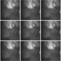
As technology continues to advance, more work is being done in the field of plaque characterization. So far all the invasive and noninvasive modalities are geared toward the detection of severely stenotic plaques (equal to or greater than 70% stenosis). However, the majority (about 70%) of acute coronary events occur in lesions with less than 50% stenosis (vulnerable plaques).1 These vulnerable plaques are characterized by a large lipid core, thin fibrous cap, and high degree of inflammation (large number of macrophages). Some early information about plaque characterization came from studying carotid arteries since they are bigger in size and with fewer motion-related artifacts compared to the heart. Using positron emission tomography (PET) images co-registered with computed tomography (CT) scan images, Rudd et al. reported that atherosclerotic plaque inflammation can be imaged with 18FDG-PET, and that symptomatic, unstable plaques accumulate more 18FDG than asymptomatic lesions.2


Stay updated, free articles. Join our Telegram channel

Full access? Get Clinical Tree


