Thoracic Aortic Aneurysm
(1,
2)1. Localized dilatation of the thoracic aorta greater than 50% of normal. The upper limit of normal caliber for the descending thoracic aorta is 3 to 3.5 cm.
2. Incidence—5.9 to 10.4 cases per 100,000 person-years
3. Male-to-female ratio—1.5:1 to 1:1
4. Classification
a. Anatomic site: 30% to 40% involve the descending thoracic aorta.
b. Morphology: fusiform (80%) or saccular (20%)
5. Risk factors—Most are “degenerative,” a late stage of atherosclerosis. Other risk factors include dissection, infection, inflammatory aortitides (e.g., Takayasu disease), connective tissue diseases (e.g., Marfan syndrome), trauma, and iatrogenesis.
6. The natural history is progressive expansion. Risk of rupture, usually fatal, markedly increases when the diameter exceeds 6 cm.
7. Conventional treatment is open surgical repair. With modern techniques, shortterm death rates range from 3% to 15% for elective cases and up to 50% for emergent operations. The risk of spinal cord ischemia is 3% to 8%.
Aortic Dissection
(1,
2,
3)1. Begins with a tear of the aortic intima and inner layer of the media, allowing blood to cleave a longitudinal plane within the media. The resulting dissection flap separates the aorta into true and false lumina. The false lumen may compress the true lumen or obstruct aortic branch vessel flow, causing end-organ ischemia. The vast majority of primary entry tears originate just distal to the left subclavian artery (
LSCA) origin or within a few centimeters of the aortic valve. Additional communications between true and false lumina may form.
2. Incidence—2 to 3.5 and 0.5 to 2.1 cases per 100,000 person-years for all dissections and those limited to the descending thoracic aorta, respectively
3. Male-to-female ratio—2:1 to 5:1
4. Classifications
a. Anatomic site
(1) Stanford system
(a) Type A—involves the ascending aorta (60% to 70%)
(b) Type B—confined to the descending thoracic aorta (30% to 40%)
(2) DeBakey system
(a) Type I—involves the ascending and descending aorta
(b) Type
II—confined to the ascending aorta
(c) Type III—confined to the descending aorta without (IIIa) or with (IIIb) extension into the abdominal aorta
b. Time from symptom onset
(1) Acute—<2 weeks
(2) Subacute—2 to 6 weeks
(3) Chronic—>6 weeks
c. Clinical course: At initial presentation, 30% to 42% are classified as “complicated” due to aortic rupture or impending rupture, malperfusion of visceral or peripheral arteries, rapid false lumen aneurysmal growth, refractory hypertension, or intractable pain.
5. Risk factors—The majority of patients have hypertension (70% to 90%). Other risk factors include existing aneurysm, connective tissue diseases (e.g., Marfan syndrome), bicuspid aortic valve, pregnancy, trauma, and iatrogenesis.
6. The natural history is highly variable. The primary late complication is false lumen dilatation and eventual rupture. Aneurysmal degeneration of the false lumen occurs in 20% to 50% within 4 years despite optimal medical therapy (OMT). Predictors of degeneration include a primary entry tear ≥10 mm, total aortic diameter ≥40 mm, false lumen diameter ≥22 mm, and partial false lumen thrombosis.
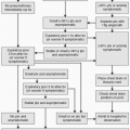
7. Conventional treatment
a. Uncomplicated type B aortic dissection (AD): OMT with control of blood pressure (goal systolic blood pressure 100 to 120 mm Hg), heart rate (goal <60 beats per minute), and pain. Lifelong close surveillance is required to
monitor for signs of disease progression, false lumen aneurysmal dilatation, and/or malperfusion.
b. Complicated type B AD: Open repair, usually performed emergently, is associated with a surgical mortality rate of 25% to 50% with paraplegia occurring in 7% to 36%. Concomitant OMT is required. Prior to the advent of thoracic endovascular aortic repair (
TEVAR), fenestration of the dissection flap to equalize pressures in the true and false lumina or stent placement to open obstructed branch vessels were the primary endovascular treatments for malperfusion (
4).
Stent Grafts
After the first thoracic aortic stent graft was implanted in a patient in 1992, the first commercial device was approved by the U.S. Food and Drug Administration (
FDA) in 2005. As of this writing, there are four FDA-approved commercial thoracic endografts (
Table 16.1), most of which are second-generation devices: Conformable TAG (W.L. GORE and Associates, Flagstaff, AZ), Valiant (Medtronic, Santa Rosa, CA), Zenith TX2 Pro-Form (Cook Medical, Bloomington, IN), and RELAY (Bolton Medical, Sunrise, FL). Other commercial devices are available outside of the United States.