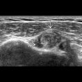
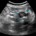
GROSS ANATOMY
Thyroid Gland
- •
H- or U-shaped gland in anterior cervical neck formed from 2 elongated lateral lobes with superior and inferior poles connected by median isthmus
- •
Thyroid gland lies anterior and lateral to trachea in visceral space of infrahyoid neck
- ○
Extends from level of 5th cervical vertebra to 1st thoracic vertebra
- ○
Posteromedially are tracheoesophageal grooves
- –
Contains paratracheal lymph nodes, recurrent laryngeal nerve, parathyroid glands
- –
- ○
Posterolaterally are carotid spaces
- –
Contains common carotid artery, internal jugular vein, vagus nerve
- –
- ○
Anteriorly are infrahyoid strap muscles
- ○
Anterolaterally are sternocleidomastoid muscles
- ○
- •
Thyroid gland
- ○
2 lateral lobes (i.e., right and left lobes)
- –
Measure ~ 4 cm in height
- –
Each lobe has upper and lower poles
- –
Lateral lobes are commonly asymmetric in size
- –
- ○
Lateral lobes are joined by midline isthmus
- ○
Pyramidal lobe present in 30-50% of cases
- –
Extends superiorly from isthmus toward hyoid bone
- –
More common on left
- –
- ○
- •
Superior thyroid arteries
- ○
1st anterior branch of external carotid artery
- ○
- •
Inferior thyroid arteries
- ○
Arise from thyrocervical trunk, branch of subclavian artery
- ○
- •
Thyroidea ima occasionally present (3%)
- ○
Single vessel originating from aortic arch or brachiocephalic artery
- ○
Enters thyroid gland at inferior border of isthmus
- ○
- •
Lymphatic drainage
- ○
Lymphatic drainage is extensive and multidirectional
- ○
Initial lymphatic drainage courses to periglandular nodes
- –
Prelaryngeal, pretracheal, and paratracheal nodes along recurrent laryngeal nerve
- –
Paratracheal nodes drain into mediastinum
- –
Regional drainage occurs laterally into internal jugular chain (levels 2-4) and spinal accessory chain (level 5)
- –
- ○
- •
Embryology
- ○
Thyroid gland forms near base of tongue (foramen cecum) and descends into neck along thyroglossal duct
- –
Ectopic thyroid gland can be seen from base of tongue into superior mediastinum
- □
Most common location is just deep to foramen cecum at tongue base (i.e., lingual thyroid)
- □
- –
Thyroglossal duct cyst results from failure of complete involution of thyroglossal duct
- –
- ○
Parathyroid Glands
- •
Small lentiform glands posterior to thyroid gland
- ○
Extracapsular in most cases but can be located within thyroid gland
- ○
- •
Located in region of tracheoesophageal groove
- •
Normal measurements
- ○
~ 6 mm in length, 3- to 4-mm transverse, and 1-2 mm in anteroposterior diameter
- ○
- •
Variable number, but typically 4
- ○
2 superior and 2 inferior
- ○
- •
Superior parathyroid glands
- ○
More constant in position as compared with lower glands
- ○
Lie on posterior border of middle 1/3 of thyroid in 75%
- ○
- •
Inferior parathyroid glands
- ○
More variable in location
- ○
50% of inferior glands lie lateral to lower pole of thyroid gland
- ○
15% lie within 1 cm of inferior thyroid poles
- ○
35% position is variable, residing anywhere from angle of mandible to lower anterior mediastinum
- ○
ANATOMY IMAGING ISSUES
Thyroid Gland
- •
Use high-resolution linear array transducer (10-15 MHz)
- •
Normal thyroid parenchyma has fine, uniform echoes and is hyperechoic compared to adjacent muscles
- •
Echogenic thyroid capsule is clearly visualized and helps to differentiate thyroid lesions from extrathyroidal masses
- •
Both longitudinal and transverse scans are required for comprehensive ultrasound assessment of thyroid gland
- ○
Measurements taken in all 3 planes
- ○
- •
Midline transverse view for isthmus
- •
Document size of all nodules with particular attention to sonographic characteristics (all features are listed from least concerning to most concerning for malignancy)
- ○
Composition : Cystic, spongiform, mixed cystic and solid, solid, or predominantly solid
- ○
Echogenicity : Anechoic, hyperechoic, isoechoic, hypoechoic, very hypoechoic
- ○
Shape : Wider than tall, taller than wide
- ○
Margin : Smooth, ill-defined, lobulated or irregular, extrathyroid extension
- ○
Echogenic foci : Comet-tail artifacts, macrocalcifications, peripheral (rim) calcifications, punctate echogenic foci
- ○
- •
Assess adjacent structures (including trachea, esophagus, strap muscles, carotid artery, and internal jugular vein) and cervical lymph nodes
Parathyroid Glands
- •
Normal glands often not seen
- ○
Look carefully in tracheoesophageal groove behind mid- to lower thyroid
- ○
Small, well-circumscribed, hypoechoic nodules posterior to thyroid gland separated by echogenic thyroid capsule
- ○
- •
Best 1st examination for localizing parathyroid adenoma
- ○
May be in ectopic location so start above thyroid at angle of mandible and move downward, scanning through thyroid to level of clavicle
- ○
Hypoechoic on grayscale with hypervascularity on color flow imaging
- –
Color flow imaging best done in longitudinal plane
- –
- ○
- •
Easy to confuse with other structures, especially cervical lymph nodes
- ○
Look for echogenic hilum and classic central hilar vessel in lymph node
- ○
THYROID AND PARATHYROID GLANDS