Trauma Management
Brian F. Stainken
Worldwide, the leading cause of death in patients younger than the age of 44 years is trauma. In 2013, trauma claimed over 2.1 million years of potential life with direct costs of over $700 billion in the United States alone. These staggering numbers do not take into consideration the additional expense of disability or the lost financial productivity of survivors (1). The cost to society of lost productivity alone is approximately four times the direct medical expenses (2).
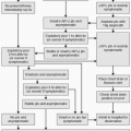
Great strides have been made toward improving trauma care in the United States. Integrated systems that initiate resuscitation at the site and expedite transfer to specialized centers have helped save lives. And these systems continue to evolve. In the past, the trauma patient was best cared for in a trauma operating room (OR). A primary goal of trauma care was to expedite transfer from the field to the OR where diagnostics and definitive care could be provided. Over the past decade however, primarily due to the deployment of helical computed tomography (CT) in the trauma bay, the path followed by the trauma patient now detours through CT. Diagnosis and triage have moved from the OR to the CT scanner. Following CT diagnosis, expedited directed care based on imaging findings follows—orthopedic, neurosurgical, laparotomy, or interventional radiology (IR) embolotherapy. Resuscitation moves with the patient. Whole-body CT scanning prior to intervention in hemodynamically unstable patients and patients requiring emergency bleeding control is associated with significantly better survival than direct transfer to surgery on arrival (3). Even the distance between the trauma bay and the CT machine independently predicts survival (4).
IR has evolved in parallel since 1972 when Margolies et al. first described embolization therapy for management of pelvic trauma. Techniques and devices to manage trauma victims nonoperatively have been validated, and the scope of care provided has expanded. IR has become an indispensable member of the trauma team, with the capability to rapidly address exsanguinating hemorrhage throughout the body without the inherent delay and morbidity of open exploration. The old bromide that unstable exsanguinating trauma patients go to the OR and the stable ones to IR no longer applies. When the IR space is appropriately configured and staffed with an experienced, trained team, the decision for IR versus OR should be based on diagnosis, not acuity. Leading trauma centers increasingly recognize the importance of image-guided medicine and the need to provide the resources and expert practitioners able to effectively integrate IR in to the trauma care environment.
The initial steps of intervention for trauma are similar regardless of the location of injury. General considerations are discussed first. The details involved in treating specific types of traumatic injury—abdominal including splenic, hepatic, and renal; pelvic; lumbar artery; and extremity are then discussed, along with their complications.
General Considerations in Trauma
Indications
1. Treatment of exsanguinating hemorrhage Although some trauma guidelines still recommend that hemodynamically unstable patients go directly to surgery, it is increasingly clear that the use of helical CT offers a major benefit for diagnosis prior to laparotomy especially in the most acute patients. As trauma centers accept this shift, and the point of diagnosis moves from the OR suite to the CT scanner, it becomes increasingly feasible to transfer directly from CT to IR in centers appropriately trained and
equipped. In fact, the time to start of emergency bleeding control procedures in one center is 45 minutes for the OR and 54 minutes for IR (3). In this changing environment, the eligibility of a patient for IR care is driven as much by the preparation and layout of the trauma facility as it is by the procedure.
equipped. In fact, the time to start of emergency bleeding control procedures in one center is 45 minutes for the OR and 54 minutes for IR (3). In this changing environment, the eligibility of a patient for IR care is driven as much by the preparation and layout of the trauma facility as it is by the procedure.
Contraindications
Relative
1. Pregnancy. A relative risk assessment may demonstrate that the morbidity of surgery exceeds the theoretical risk of radiation exposure especially in late trimester gestation. Advanced planning for such scenarios is paramount.
2. Renal insufficiency. Acute tubular necrosis related to prolonged hypotension is a much more common issue in trauma patients than contrast nephropathy.
3. Allergy to contrast. Clarification of the actual history (idiosyncratic vs. anaphylactic) is important. In most patients, this issue will be resolved at the time of CT imaging. Depending on the presentation and relative risk, even in a known allergic patient, it may be reasonable to proceed after consultation with other members of the trauma team.
Preprocedure Preparation
1. Ongoing resuscitation is critical during all aspects of trauma care including endovascular intervention. This entails ensuring an airway and appropriate ventilation as well as adequate intravenous (IV) access with large-bore IV lines for fluid, medication, and transfusions. Warming the patient and replacement of coagulation factors will aid in hemostasis. Other than cervical stabilization, neurologic assessment can wait in hemodynamically labile patients. Although associated with a poorer prognosis, neurologic injury alone is not likely to cause significant hemodynamic shock.
2. Basic laboratory studies are not a prerequisite for trauma angiography, and the procedure should not be delayed in anticipation of complete blood count (CBC), chemistry panel, and coagulation parameters. A worsening base deficit is the most sensitive laboratory indicator of hemodynamic shock and will precede a drop in hematocrit.
3. Review of CT and other available imaging studies to identify all potential sites of injury and occult sources of bleeding is essential in formulating a plan for intervention. The trauma CT is the cornerstone of initial diagnosis. A negative CT in children is 99.6% predictive to exclude any intra-abdominal injury (5). In penetrating abdominal trauma, CT has far greater sensitivity than focused assessment with sonography for trauma (FAST) and emerging data suggest that its use as a primary tool (during resuscitation) saves lives (3,6).
4. Resuscitation should be continued throughout the procedure. A lifesaving procedure should not be postponed while waiting for blood products. Safe embolization in unstable patients can be performed if the patient responds, even transiently, to a 2-L rapid resuscitative bolus (7). Transfusions should be administered while the patient is in the angiography suite to prevent delays.
Procedure
1. Arterial access is usually obtained via the common femoral artery. A vascular sheath is essential in all cases. In the setting of severe hemodynamic shock, the femoral pulses may not be palpable, and ultrasound should always be available for access. Upsize the sheath 1 to 2 Fr. beyond the planned catheter size, and use the side arm connected to a manometer to monitor arterial pressures. The same sheath can be used, when warranted for resuscitation. Sew the sheath in if you expect it to be left in place after your procedure.
2. Diagnostic angiographic images have largely been supplanted by the CT scan. Treatment should not be delayed for the purpose of angiographic documentation. If extravasation is seen on the hand-injected fluoroscopic test imaging,
save it and move on to treatment without a formal injector run when the situation calls for speed. Angiographic findings for vessel disruption in the setting of trauma may include extravasation, occlusion, arteriovenous (AV) or other fistula, intimal tear, and false aneurysm. Extravasation seen on CT and not seen at angiography may be due to vessel spasm, slow bleeding rate (e.g., venous bleeding), changes in blood pressure, cardiac output, or anomalous anatomy. Remember to always obtain orthogonal views when the finding is not clear in one projection. A single angiographic projection can miss significant findings and should not be relied on to clear a vessel of injury. Selective runs may show extravasation not visible on a flush run.
save it and move on to treatment without a formal injector run when the situation calls for speed. Angiographic findings for vessel disruption in the setting of trauma may include extravasation, occlusion, arteriovenous (AV) or other fistula, intimal tear, and false aneurysm. Extravasation seen on CT and not seen at angiography may be due to vessel spasm, slow bleeding rate (e.g., venous bleeding), changes in blood pressure, cardiac output, or anomalous anatomy. Remember to always obtain orthogonal views when the finding is not clear in one projection. A single angiographic projection can miss significant findings and should not be relied on to clear a vessel of injury. Selective runs may show extravasation not visible on a flush run.
3. Options for achieving hemostasis when faced with a traumatic vascular injury include embolization, stent or stent-graft placement, and temporary balloon occlusion.
a. Embolization may be performed using gelfoam slurry or by selectively delivering compressed dry pledgets aptly called torpedoes. One may employ particulate microemboli such as polyvinyl alcohol (PVA) or spherical embolics, macroembolic metallic coils, endoluminal plugs, or any combination of these. Gelfoam is commonly used in situations where rapid, temporary vascular occlusion is needed. It is well suited for use in occluding small end arteries and when faced with diffuse branch bleeding as is often seen in the setting of pelvic fractures. Coils are generally used for embolization of larger conduit vessels or to isolate arterial fistulas. Detachable coils and plugs allow precise control over delivery.
b. Stents and stent grafts can rapidly control exsanguinating bleeding while preserving luminal continuity when larger conduit vessels such as the aorta, carotid, hepatic, renal, iliac, and superficial femoral arteries are disrupted.
c. Temporary balloon occlusion is an important component in the IR arsenal for hemostasis. Inflating an appropriately sized, compliant balloon within the artery proximal to a site of extravasation may provide immediate hemostatic control. The central lumen can then be used to better study the bleeding site and plan care. Microcatheters can be deployed through the central lumen to deliver emboli if indicated. Downstream pressures can be measured. Temporary balloon occlusion can achieve proximal control during surgical exposure and preemptive balloon placement may be a useful adjunct when there is high potential risk for bleeding (8). One example is the use of temporary balloon occlusion in penetrating subclavian artery injury for proximal hemostatic control and repair via a clavicular resection rather than sternotomy.
4. Once stabilized, completion angiography is important to confirm hemostasis prior to removal of the access sheath. Closure devices should be considered in this population as patients may be heavily sedated, may proceed for additional procedures, and/or be at high risk for transfusion-related coagulopathy.
Postprocedure Management
1. All trauma patients should be followed closely for signs of persistent or delayed hemorrhage with serial blood work, physical exam, and vital sign checks. It is the responsibility of the treating physician to identify complications and ensure that the patient has fully recovered from the intervention.
2. Adequate prophylaxis for infection and deep vein thrombosis (DVT) should be considered in trauma patients.
3. Follow-up angiography after endovascular treatment of arteriovenous fistulas (AVFs) is recommended.
Abdominal Trauma
In order of frequency, the most commonly injured abdominal organs are the spleen, liver, and kidney. With the widespread use of cross-sectional imaging and
endovascular techniques, the majority of hemodynamically stable or adequately resuscitated abdominal trauma patients, can be nonoperatively managed.
endovascular techniques, the majority of hemodynamically stable or adequately resuscitated abdominal trauma patients, can be nonoperatively managed.
Splenic Injury
Historically, traumatic injury to the spleen was treated with splenectomy or observation. With the growing acceptance of splenic artery embolization, most patients can safely avoid surgery (9). The immunologic benefits of splenic preservation are accepted (10). There remains great variability in practice even between major trauma centers. One comparative review of four level 1 trauma centers showed that centers with the highest rates of embolization also enjoy the highest rates of splenic salvage (11). The probability of successful preservation of the spleen is increased when embolization is used for all grade 3 and 4 lacerations (12).
Indications
1. Blunt trauma
a. Extravasation of contrast noted on arterial phase CT or on angiography. There is some debate as to the merit of embolization over observation for stable patients presenting with a grade 1 to 3 laceration and parenchymal contrast extravasation “blush” on CT (13).
b. Hemodynamic lability or transfusion requirements greater than 4 units packed red blood cells (PRBC) over 24 hours. Current surgical society standards advise against embolization in the setting of hemodynamic instability; however, larger series demonstrate clear shifts in the percentage of patients undergoing embolization without a change in the distribution of splenic injury scores (9).
c. CT findings of splenic injury of grade 3 (laceration >3 cm or >5 cm hematoma) or vascular (involvement of segmental or hilar vessel) laceration with or without active extravasation. Segmental infarction of the spleen noted on CT


Related posts:


Stay updated, free articles. Join our Telegram channel
Full access? Get Clinical Tree
