Abstract
Tri-orchidism which means 3 testes in the scrotal cavity, is the most common type of the polyorchidism. It is the rarest anomaly of the urogenital tract, and occurs more commonly on the left side. Superanumerary testes shows scrotal and extra-scrotal presentation and is associated with several clinical features and complications. We report a case of triorchidism in a 29-year-old man who presented with left hemiscrotal pain, later on diagnosed on clinical and sonographic examinations. The management depends on different factors and include conservative treatment with follow up, orchidopexy and orchidectomy. Orchidectomy is advised in suspected case of malignancy.
Introduction
The term polyorchidism means more than 2 testes in either hemiscrotum. It is an extremely rare congenital anomaly of urogenital tract. The most common type is tri-orchidism where 3 testes are present. In literature, less than 200 cases are reported [ ]. The first case of triorchidism was reported by Blasius in 1670 at routine autopsy [ ]. The exact cause is not known, but some suggest that during the early embryogenesis, the longitudinal or transverse division of the urogenital ridge by peritoneal bands development is the possible mechanism. This entity is incidentally diagnosed during surgical exploration [ ].
Tri-orchidism occurs more on the left side, because the left testis is more prone to subdivision due to its larger size and different vascular anatomy as compared to the right testis [ , ]. The extra testis is commonly present in the Scrotum (66%), inguinal canal (23%) or retroperitoneum (9%) [ ].
Polyorchidism is associated with several complications including maldescent (40%), inguinal hernia (30%), malignancy (6%), hydrocele (9%), infertility, testicular torsion and varicocele [ ].
Management of patients with triorchidism is controversial and depends on multiple factors including the location of the testis, the reproductive potential, size of the testis and age of the patient [ ].
Here we report a case of tri-orchidism in a 29-year-old man with imaging features on ultrasound and MRI, who incidentally diagnosed during scrotal ultrasound scan for pain.
Case report
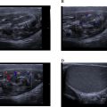
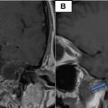
A 29-year-old male presented to urology department of our hospital, who has been complaining of nontraumatic dull pain in left hemiscrotum. On physical examination the left hemiscrotum revealed a tender round lesion superior to the left testis which was firm and mobile, while right testis was normal. There was no evidence of lymphadenopathy or inguinal hernia. He was referred to radiology department for ultrasound. On sonography, the left hemiscrotum demonstrated 2 small sized testicles with homogeneous echo-texture and normal blood flow, each having equal volume of 6 cc and 5.8 cc respectively. They showed shared epididymis and vas deference ( Fig. 1 ). There was no evidence of hydrocele, varicocele or any other pathology. The right testis was normal and larger than both testes in left hemiscrotum. The complementray MRI revealed that the left hemiscrotum containing 2 round to oval shaped structures with signal characteristics identical to normal testicular tissue ( Fig. 2 ). The man was diagnosed with type II polyorchidism and referred back to the urologist, who decided to manage the patient conservatively considering the patient’s age and need to maintain the fertility, and advised the patient for routine follow up with instructions to report any changes in size, shape, or pain.


Related posts:
 Breast cancer with medullary features shows a fast and plateau enhancement pattern on magnetic resonance images: A case report
 A rare and life-threatening case of spontaneous hemopneumothorax presenting with severe hemodynamic instability
 Avascular necrosis of lateral femoral condyle in a middle-aged woman from central India
 A very rare case of ileocolic and appendiceal intussusception with acute appendicitis
 Rare case of left epididymo-orchitis complicated by pampiniform plexus thrombosis: A case report
 Petrous apex epidermoid cyst: A rare case
Stay updated, free articles. Join our Telegram channel

Full access? Get Clinical Tree


