KEY FACTS
Imaging
- •
Holoprosencephaly is hallmark anomaly
- ○
Spectrum of severity: Alobar, semilobar, lobar
- ○
Associated facial anomalies
- –
Close-set orbits (hypotelorism), fused eyes (cyclopia)
- –
Small nose, proboscis (tube-like and above orbits)
- –
Cleft lip/palate: Midline or bilateral
- –
- ○
- •
Cardiac defects in most cases
- •
Large echogenic kidneys
- •
Postaxial polydactyly: Extra finger or toe next to 5th digit
- •
Early fetal growth restriction commonly seen
- •
Detectable at time of nuchal translucency (NT) screening
- ○
↑ NT, absent nasal bone, holoprosencephaly
- ○
Top Differential Diagnoses
- •
Holoprosencephaly without trisomy 13: Similar facial anomalies
- •
Trisomy 18: Also associated with holoprosencephaly
- •
Meckel-Gruber: Encephalocele, cystic kidneys, polydactyly
Clinical Issues
- •
Advanced maternal age at higher risk
- •
3rd most common trisomy (after trisomy 21 and T18)
- •
Offer genetic testing when hallmark anomalies seen
- ○
Chorionic villus sampling, amniocentesis
- –
Cell-free fetal DNA is for screening only
- –
- ○
- •
Prognosis: 50% die in utero, 80% of live-born die 1st day of life, < 10% reach 1st birthday
Scanning Tips
- •
Look for falx at time of NT scan: Absent falx with fluid crossing midline suggests monoventricle of holoprosencephaly
- •
Profile view showing premaxillary protrusion at time of NT scan or midgestation is highly suggestive of bilateral cleft lip/palate
- •
If cavum septum pellucidum is absent, look for fused frontal horns (may be only finding of lobar holoprosencephaly)
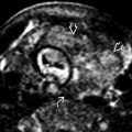
 and thin anteriorly fused brain mantle
  across the midline. The thalamus is fused and “ball-like”
  , classic findings of holoprosencephaly. The NT was also increased in this fetus.
 . The falx is absent and the lateral ventricles are fused and cross the midline
  . Part of a proboscis
  is also seen.
 caused by forward displacement of the anterior palate and soft tissue from bilateral cleft lip and palate. An otherwise normal nasal bone
  and chin
  are seen in this case.