Tumors of the Pineal Region
Overview
Primary tumors of the pineal region are comprised largely of four different histologic subtypes—pineocytoma, pineal parenchymal tumor of intermediate differentiation (PPTID), pineoblastoma, and papillary tumor of the pineal region (PTPR). Each has unique cell of origin, gender and age predilection, imaging features, and prognosis.
| ||||||||||||||||||||||||||||||||||||||||||
Tumor Types
Pineocytoma
Pineal parenchymal tumor of intermediate differentiation (PPTID)
Papillary tumor of the pineal region (PTPR)
Pineoblastoma
Pineocytoma
Definition: Pineocytoma is a well-differentiated pineal parenchymal tumor comprised of uniform cells forming large pineocytomatous rosettes and/or of pleomorphic cells showing gangliocytic differentiation. It is a WHO grade I tumor with no malignant potential or dissemination.
Epidemiology: This accounts for <1% of all intracranial neoplasms and 27% of pineal neoplasms.
Affected age group: The tumor can occur in any age but most frequently affects adults, with a mean age of 43 years; there is a female predominance, with a female-to-male ratio of 1:0.6.
Molecular and genetic profile: Cell of origin is likely the pinealocyte, a cell with photosensory and neuroendocrine functions. It is associated with immunoexpression of cone-rod homeobox (CRX) (a transcription factor involved in the development and differentiation of pineal cell lineage) and acetylserotonin methyltransferase (ASMT) (a critical enzyme for synthesis of melatonin, which is a hormone produced by the pineal gland). No syndromic associations or genetic susceptibilities are known.
CSF markers: None.
Clinical features and standard therapy: Surgical resection is the definitive treatment without adjuvant radiotherapy or chemotherapy.
Prognosis: The 5-year survival rate ranges from 86% to 91%, and the most important prognostic marker is the extent of surgical resection.
Imaging
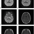
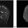
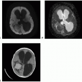
The hallmark of pineocytoma is a lobulated solid mass with or without cystic or calcific component, usually <3 cm, which may cause obstructive hydrocephalus. The cystic component may be the dominant feature with minor solid tumor tissue.
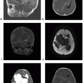
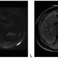
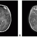
 Figure 9.1. (continued) G. Axial T1 postcontrast: Thin rim of enhancement of the cystic mass. H. Axial T1 postcontrast: Thin rim of enhancement of the cystic mass. I. Sagittal T1 post contrast: Thin rim of enhancement of the cystic mass.
Stay updated, free articles. Join our Telegram channel
Full access? Get Clinical Tree
 Get Clinical Tree app for offline access

|

