Tumors of the Sellar Region
Overview
The genetic underpinnings of adamantinomatous and papillary craniopharyngiomas are very different, suggesting that they are each a distinct tumor entity although they share the same name. On the contrary, the three rare tumors of the sellar region—granular cell tumor, pituicytoma, and spindle cell oncocytoma—are thought to arise from multiple subtypes of pituicytes, hence sharing similar genetic expression.
|
Adamantinomatous Craniopharyngioma
Definition: Adamantinomatous craniopharyngioma is a benign, cystic and solid, epithelial tumor arising from embryonic remnants of the Rathke pouch epithelium.
Epidemiology: This tumor accounts for 1.2% to 4.6% of all intracranial tumors with bimodal age distribution.
Molecular and genetic profile: Catenin beta 1(CTNNB1) mutations and aberrant nuclear expression of beta-catenin in up to 95% of cases are seen.
Clinical features and standard therapy: Prominent cystic components of adamantinomatous craniopharyngioma are often multiloculated and multicompartmental, rendering complete surgical removal difficult, as is the infiltrative growth of the solid component. Recurrence is common when there is residual tumor after the initial surgery, and both chemotherapy and radiation therapies are largely ineffective.
Imaging
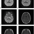
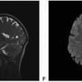
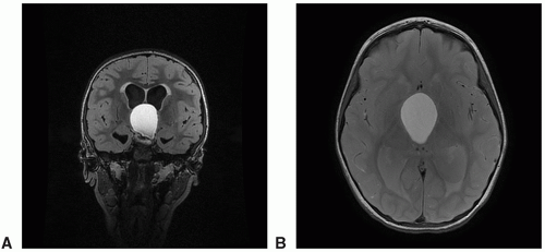 Figure 10.1. Imaging of adamantinomatous craniopharyngioma. A. Coronal FLAIR: Hyperintense cystic mass. B. Axial fluid-attenuated inversion recovery (FLAIR): Hyperintense cystic mass. |
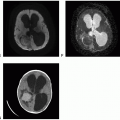
 Figure 10.2. Imaging of adamantinomatous craniopharyngioma. A. Axial FLAIR: Hyperintense cystic mass with layering blood products. B. Axial T2: Hyperintense cystic mass with layering blood products.
Stay updated, free articles. Join our Telegram channel
Full access? Get Clinical Tree
 Get Clinical Tree app for offline access

|


