Abstract
Sternal metastasis often result in disabling pain, significant functional limitations, with potential consequences for the thoracic spine. This case report suggests a new approach combining ultrasound for initial guidance and fluoroscopy with cone-beam CT (CBCT) for screw fixation. The patient experienced immediate pain relief, a better mobility, and improved quality of life. This approach demonstrates a minimally invasive, radiation-sparing and time saving strategy for sternal screwing.
Introduction
Sternal metastases, often originating from breast, renal, or lung cancer, frequently lead to painful fractures that significantly impair patient quality of life. These pathological fractures often require stabilization to alleviate pain and improve function [ ].
While surgical resection remains the standard for isolated metastases, it is often invasive and associated with postoperative complications [ ]. Minimally invasive approaches, including percutaneous screw fixation, cementoplasty, and thermal ablation techniques such as radiofrequency ablation (RFA) and cryoablation, have gained traction due to their ability to provide pain relief, stabilize fractures, and improve patient function with reduced morbidity [ ]. Despite these advancements, there is limited data on the use of ultrasound guidance in percutaneous sternal fixation, which could provide an effective alternative by reducing radiation exposure and enhancing procedural accuracy
This case report describes a novel hybrid imaging approach that integrates ultrasound for initial trocar placement, followed by fluoroscopic and CBCT confirmation, in the percutaneous screw fixation of a sternal metastatic lesion. By presenting the technical feasibility, safety, and clinical outcomes, this study aims to highlight ultrasound guidance as a valuable addition to interventional radiology techniques for sternal metastases, offering a less invasive, radiation-sparing, and time-efficient alternative to conventional methods
Case report
The patient is a 71-year-old female with a history of metastatic renal cancer, diagnosed in 2015 with clear cell renal carcinoma and multiple metastases treated with surgery and immunotherapy. She has since experienced progressive metastatic disease involving the lungs, bones, and digestive complications. Despite multiple therapeutic adjustments (including nivolumab, cabozantinib, and axitinib) and stable pulmonary lesions, bone metastases, notably in the sternum, led to severe symptoms. The blood test revealed elevated monocytes (0.78 G/L; reference: 0.17-0.56) and creatinine clearance of 52 mL/min/1.73 m 2 , suggesting mild renal impairment and a potential inflammatory or immune-related process. All other parameters, including coagulation and thyroid function, were within normal ranges. The patient presented with severe chest pain localized to the lower sternum for 3 weeks despite analgesic treatment including opiod. The patient reported severe pain upon palpation of the sternum, which worsened with movement and breathing. Preoperative CT imaging revealed a lytic lesion in the lower half of the sternum, with healthy cortical remnant of the lower end of the sternal body ( Fig. 1A ). Following a multidisciplinary team discussion with spine surgeons, radiation oncologists, medical oncologists and interventional radiologists, it was decided to proceed with percutaneous screw fixation.

The procedure was performed under general anesthesia with the patient positioned in supine position. 2 g of Cefazolin were administrated intravenously.
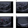
The first step involved inserting of a 22-gauge spinal needle under ultrasound guidance (Acuson Freestyle, Siemens Healthineers, Princeton, NJ) up to bone contact with the remaining intact cortex. Confirmation of the needle position was achieved using cone-beam computed tomography (CBCT) (Artis Zee, Siemens Healthineers, Princeton, NJ). Once the bone entry point was confirmed, a 1 cm incision was made 7 cm below the target area to provide access to the inferior border of the sternum. This incision enabled the safe placement of an 8-gauge trocar bone (Stryker, Kalamazoo, MI), which was advanced under ultrasound guidance, through the tumor with good visibility until it reaches the remaining healthy bone medullary ( Figs. 1B and C ). After confirmation of the trocar’s placement, the imaging modality was switched to lateral fluoroscopy and a 3.2 mm Kirschner wire ( Fig. 1D ) followed by a partially threaded titanium screw with a 6.5 mm diameter and 90 mm length (Asnis III, Stryker, Kalamazoo, MI) were inserted in the sternal body ( Fig. 2 ). Iterative CBCT imaging was used throughout the procedure to monitor the screw’s progression, ensuring precise placement within both the tumor and healthy bone while avoiding cortical breaches. The procedure was completed successfully without complications. Overall, the operating time was 40 minutes. The patient experienced immediate and total pain relief, with the Numeric Pain Rating Scale (NPRS) score decreasing from 7/10 preoperatively to 0/10 postoperatively. This was confirmed at the 1-month consultation with the interventional radiologist, with an overall improvement in the patient quality of life.

Related posts:
 Breast cancer with medullary features shows a fast and plateau enhancement pattern on magnetic resonance images: A case report
 A rare and life-threatening case of spontaneous hemopneumothorax presenting with severe hemodynamic instability
 Avascular necrosis of lateral femoral condyle in a middle-aged woman from central India
 A very rare case of ileocolic and appendiceal intussusception with acute appendicitis
 Rare case of left epididymo-orchitis complicated by pampiniform plexus thrombosis: A case report
 Hepatic and extra-hepatic hydatid cysts: A case series of radiological and clinical insights
Stay updated, free articles. Join our Telegram channel

Full access? Get Clinical Tree


