Abstract
Objective
To evaluate the correlation between liver viscosity, as measured by shear wave dispersion (SWD), and fibrosis and inflammation in patients with autoimmune hepatitis (AIH). Additionally, to assess its potential as a non-invasive biomarker for hepatic fibrosis compared to shear wave elastography (SWE).
Methods
This prospective study included 25 AIH patients who underwent pre-biopsy SWD and SWE measurements using the SuperSonic Mach30 system. Liver biopsy samples were assessed for fibrosis using the Scheuer grading system and for inflammation using the modified Hepatic Activity Index (mHAI). Correlations between viscosity, elastography, fibrosis, and inflammation were analysed using Spearman’s correlation coefficients.
Results
Viscosity demonstrated a significant correlation with fibrosis stages (Spearman’s coefficient: 0.58, p = 0.002), while SWE showed a weaker correlation (Spearman’s coefficient: 0.50, p = 0.01). Viscosity measurements also correlated moderately with the mHAI score (Spearman’s coefficient: 0.62, p = 0.001). Subgroup analyses revealed weak to moderate correlations between viscosity and mHAI components across fibrosis stages.
Conclusion
Our study suggests that viscosity may be better than SWE as a non-invasive marker for assessing hepatic fibrosis in AIH, particularly in the pre-treatment period when inflammation levels are elevated. However, we could not conclusively determine the relationship between viscosity and hepatic inflammation, as a small sample size limited our findings. Further research with a larger cohort of AIH patients is necessary to better understand the correlation between viscosity and inflammation in this rare condition.
Introduction
Autoimmune hepatitis (AIH) is a rare, immune-mediated hepatic inflammatory disease that frequently manifests as acute hepatitis but may evolve into a chronic condition, leading to liver cirrhosis and eventually end-stage liver disease [ , ]. The diagnosis of AIH can be challenging due to the overlap of symptoms with other forms of acute hepatitis and the absence of pathognomonic clinical or biochemical markers [ , ]. As of today, the definitive diagnosis of AIH relies on liver biopsy, which confirms compatible histological findings alongside supportive biochemical tests [ ]. However, liver biopsies are costly, invasive, carry a small risk of complications, and in rare instances, may result in mortality [ ]. Consequently, there is an increasing shift toward utilizing non-invasive diagnostic and follow-up methods to reduce the risks, costs, and challenges associated with liver biopsies [ ].
Two-dimensional shear wave elastography (2D-SWE) is a non-invasive ultrasound-based method that enables real-time examination of the hepatic parenchyma using focused ultrasound pulses to generate shear waves within the tissue. Shear waves are mechanical waves generated within tissues that travel perpendicular to the direction of the applied force. Because shear waves travel faster through stiffer fibrotic tissue, the speed of these waves can be used to calculate and map liver stiffness [ ]. Numerous studies have indicated that 2D-SWE is a valuable non-invasive tool for assessing liver fibrosis [ ]. Recently, a novel technique that extends the capabilities of 2D-SWE has been developed, specifically targeting the analysis of shear wave dispersion (SWD) properties. This approach focuses on the variation of shear wave velocity with frequency, a parameter known as SWD, to offer a view of organ viscosity [ ]. Viscosity, a key biomechanical property, refers to a tissue’s resistance to deformation or flow, influencing the behaviour of shear waves. When a tissue exhibits dispersion, shear wave velocity changes with frequency due to its viscous properties. This relationship allows viscosity to be approximated by analysing shear wave speed at specific frequencies. The slope of the curve plotting shear wave speed against frequency serves as an indirect measure of viscosity. SWD analysis is conducted using two technologies: Canon’s Aplio system (Japan), which features the Shear Wave Dispersion Slope (SWDS), measuring dispersion in units of (m/s)/kHz to demonstrate frequency-dependent variations in shear wave speed. A second system, developed by SuperSonic Imagine (France), utilises the Viscosity Plane Wave Ultrasound (Vi-PLUS) software integrated into the Aixplorer ultrasound system. Vi-PLUS employs ultra-fast plane-wave imaging, emitting multiple ultrasound waves at varying frequencies simultaneously to generate and track shear waves within the tissue. Advanced algorithms in the software isolate the viscous component of tissue properties from the elastic component, enabling precise quantification of tissue viscosity. The system provides a viscosity index, measured in Pascal seconds (Pa.s), offering a direct and quantitative assessment of tissue viscosity.
Beyond its ability to assess hepatic fibrosis, SWD is also regarded as a marker for parenchymal inflammation, edema, and necrosis [ , ]. Limited research, including animal studies and small-scale clinical cohorts, has demonstrated a correlation between hepatic inflammatory activity and viscosity compared with histological findings [ ]. Moreover, data from a recent retrospective study showed a decrease in SWDS in patients with AIH receiving steroid treatment, suggesting that AIH may be among the conditions demonstrating a correlation between hepatic inflammation and viscosity [ ]. Our study is the first to prospectively assess the correlation between hepatic viscosity, using SWD, and histopathologic inflammatory activity specifically in patients with AIH. This focus on AIH makes our research unique, as previous studies have primarily investigated other liver conditions or used retrospective methodologies.
Materials and methods
This single-center, prospective study was conducted at Emek Medical Center’s liver clinic, an academic-affiliated hospital. It received institutional review board approval, aligning with the ethical standards set by the committee on human experimentation and the principles of the Declaration of Helsinki.
Between August 2021 and September 2024, patients with suspected AIH who were scheduled for liver biopsies were recruited for the study. The eligibility criteria included patients over 18, with a baseline platelet count above 80,000 per cubic millimeter and a prothrombin time with an international normalised ratio of ≤1.5. After receiving a comprehensive explanation of the study’s aims and procedures, all participants provided signed informed consent in the presence of a physician.
The study was initiated following a software upgrade to the SuperSonic Mach30 sonar device (SSI, France), which enables the assessment of liver viscosity. On the day of recruitment, the patients underwent a sonographic SWE and SWD examination, using the enhanced SuperSonic Mach30 device, which included a Vi-PLUS test to evaluate liver viscosity. The examination was conducted before the biopsy as part of the diagnostic assessment. To ensure optimal test conditions, participants were advised to fast for 4 hours before the examination, with unrestricted water intake, adhering to standard biopsy preparation protocols.
Sonographic examinations were conducted by a single trained radiologist at the diagnostic imaging institute using a C6-1 convex transducer with frequencies ranging from 1 to 6 MHz. Shear wave velocity was measured and expressed as kPa. Liver viscosity was measured and expressed in Pa.s. Measurements were performed on the right lobe of the liver using an intercostal approach. The SWE and SWD area measured was at least 1 cm deep to the liver capsule and free of other anatomical structures, such as blood vessels or bile ducts. After confirmation of an appropriate SWE and SWD map, patients were asked to hold their breaths while the image was saved. A reliable result was defined as the median value of at least five measurements of SWE and SWD with a Stability Index of > 90%.
Following sonographic examination, the patients underwent liver biopsy. Using a 16 gauge automatic biopsy gun (Merit Medical USA), we performed percutaneous liver biopsies through an intercostal space targeting segments V or VIII, aiming for precision and minimal complications. Biopsies were strategically performed near the regions assessed by 2D-SWE. From each participant, 2–3 liver specimens were successfully extracted, each approximately 2 cm in length, to closely correlate histological evaluations with elastography findings. The obtained samples were then dispatched for histopathological analysis. The material was processed and embedded in paraffin, sectioned, and stained with hematoxylin and eosin using a Ventana HE600 machine (Roche USA) and then moved to the pathologist’s table. Histopathological assessment for was performed using the Modified Hepatic Activity Index (mHAI) grading system. This system quantifies necroinflammatory activity based on a numerical scoring of four key variables: A- periportal or periseptal interface hepatitis (scored from 0 to 4); B- confluent necrosis (scored from 0 to 6); C- focal necrosis/apoptosis and focal inflammation (scored from 0 to 4); D- portal inflammation (scored from 0 to 4). The scores from these four variables were summed to derive the total mHAI score, ranging from 0 to 18, indicating the overall necroinflammatory activity grade within the liver tissue [ ]. Fibrosis stages were assessed using the Scheuer grading system [ ]. Moreover, the levels of liver enzymes, aspartate aminotransferase (AST) and alanine aminotransferase (ALT), as well as immunoglobulin G (IgG) levels, were extracted from medical records spanning the three months leading up to the biopsy date. These tests were not conducted on the day of the biopsy. Finally, the diagnosis of AIH was determined using the Simplified Criteria for the Diagnosis of Autoimmune Hepatitis [ ].
Variables were collected from patients’ records and compiled into an Excel table as encoded data. Categorical variables are presented with counts and percentages, and continuous/ordinal variables are presented with median and range due to the small sample size. Spearman correlation analysis was conducted to investigate the relationship between the viscosity and elastography levels and the mHAI score. The analysis was also used to examine correlations between fibrosis levels and liver stiffness (LS), as well as between fibrosis levels and viscosity. Non-parametric local regression (LOESS smoothing) was used for the presentation (with 95% CI) of the association trend. Associations between fibrosis levels and study variables were assessed using the Kruskal-Wallis test for continuous/ordinal variables and Fisher’s exact test for categorical variables. Statistical analyses were performed using SAS 9.4 software (SAS Institute Inc., Cary, NC, USA).
Results
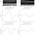
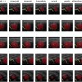
Our study initially enrolled 30 patients, of whom 25 were included in the final analysis. One participant was excluded due to human error of failure to obtain viscosity measurements before their biopsy. Four other patients who did not have a final diagnosis of AIH were excluded. All other patients were conclusively diagnosed with AIH. Figure 1 and 2 demonstrate examples of sonographic viscosity measurements and pathological images depicting the different criteria of the mHAI score of two participants from the study.


Table 1 presents the characteristics of the study participants, including measurement values, firbrosis and inflammatory scoring. The Spearman correlation between fibrosis and viscosity demonstrated a medium correlation of 0.58 ( p = 0.002). The Spearman correlation between fibrosis levels and elastography was 0.50 ( p = 0.01). The scatterplot with LOESS smooth for trend, demonstrated in Figure 3 , illustrates these two correlations. The Spearman correlation between SWE and viscosity was 0.92 ( p = 0.0001).
| Participants | |
|---|---|
| N=25 | |
| Age (years) | 58 [24–81] |
| Age≥60 | 11 (44%) |
| Gender—females | 21 (84%) |
| Elastography (kPa) | 10.1 [5.4–37.2] |
| Viscosity (Pa.s) | 2.2 [1.7–4.3] |
| Fibrosis | |
| 0–1 | 9 (36%) |
| 2 | 5 (20%) |
| 3 | 8 (32%) |
| 4 | 3 (12%) |
| Modified HAI Score | 7.0 [6.0–8.0] |
| A | 3.0 [2.0–3.0] |
| B | 0 |
| C | 3.0 [2.0–3.0] |
| D | 2.0 [1.0–3.0] |
| Modified HAI Score≤4 | 2 (8%) |
Related posts:
 Quantitative Analysis of Contrast-Enhanced Ultrasound Images of Brain-Dead Donor Livers: Prediction of Early Allograft Dysfunction
 Safety of Shear Wave Elastography as Evidenced From Carotid Artery Strain and Strain Rate Induced by Acoustic Radiation Force Impulse and Arterial Pulsations
 SegFormer3D: Improving the Robustness of Deep Learning Model-Based Image Segmentation in Ultrasound Volumes of the Pediatric Hip
 Diagnosis of Salivary Gland Tumors Using Ultrasound Radiomics
 Clutter-Generating Phantom Material. Part II: Utilization in the Comparison of Conventional and Regularized Ultrasound Attenuation Estimation
 Low-intensity Pulsed Ultrasound Stimulation of the Intestine Improves Insulin Resistance in Type 2 Diabetes
Stay updated, free articles. Join our Telegram channel

Full access? Get Clinical Tree


