Abstract
Objective
This study aimed to develop, validate and test the clinical feasibility of ultrasound (US) speckle tracking method based on gradient optical flow for quantifying small longitudinal displacements, shear and strain in peripheral nerves.
Methods
The speckle tracking method was validated using seven thawed, fresh-frozen isolated cadaveric forearms. Longitudinal motion of the median nerve was captured using a high-frequency 22 MHz linear probe. An air bubble marker was inserted as a reference point for manual measurement comparison. The precision and accuracy of the method were assessed by comparing manual and automatic measurements. Clinical feasibility was tested on eight healthy subjects, measuring the longitudinal displacement of the median nerve during elbow extension and shoulder anteflexion.
Results
The method demonstrated linearity, high precision and accuracy, particularly with a backtrace of five frames, reducing the displacement underestimation to 4%. In cadaveric models, the highest shear strain was observed at the nerve-tissue interfaces. In healthy subjects, the mean displacement of the median nerve was 3.3 ± 1.0 mm, with good inter-rater reliability (intraclass correlation coefficient = 0.87).
Conclusion
The US speckle tracking method based on gradient optical flow effectively quantifies small longitudinal displacements and shear strain in peripheral nerves, with high precision and accuracy. However, the method could not detect longitudinal strain in nerves within the range of tested displacements. Future studies should investigate its applicability to smaller and deeper nerves and its usefulness in different pathological conditions.
Introduction
With advances in high-resolution ultrasound (US) technology and the utilization of high-frequency probes (20 MHz and above), nerve US has emerged as the gold standard for imaging peripheral nerves. This is primarily attributed to its excellent spatial resolution and the capability for dynamic imaging [ ]. Because changes in the structural dispositions and biomechanics of peripheral nerves and surrounding tendons are known to correlate with the clinical severity of various entrapment neuropathies (i.e., carpal tunnel syndrome – CTS), US imaging has become an increasingly important tool for confirming clinically suspected entrapment neuropathies [ , ]. In addition to static US imaging, a few studies have utilized dynamic US imaging in patients with CTS, demonstrating the potential of dynamic assessment of the median nerve in these patients [ ]. These studies assessed either transverse or longitudinal mobility of the median nerve at the wrist. The transverse mobility of the median nerve has been shown to be reduced in patients with CTS. However, due to the pathophysiological theory underlying this pathology, transverse mobility may not be as characteristic as longitudinal mobility. Few studies have investigated the possibility of using longitudinal mobility of the median nerve to increase the diagnostic accuracy of US examination in CTS. While these studies generally indicated a decrease in the longitudinal mobility of the median nerve at the wrist, the findings across the studies have been inconsistent [ , ]. These inconsistencies can be attributed to methodological variations and technical considerations when analysing longitudinal displacement using US videos.
Researchers have developed many different methods to estimate longitudinal nerve movement, including Doppler US [ ], speckle tracking with cross-correlation [ ] and sum-absolute-difference [ ] or velocity vector algorithms [ ]. Cross-correlation methods are frequently utilized to track specific speckles within a restricted number of regions of interest inside the nerve tissue, primarily quantifying gross nerve movement [ , ]. These methods operate under the assumption that the nerve is a homogeneous structure undergoing movement as a single entity, thereby precluding the tracking of individual nerve layers. However, the nerve structure is not homogeneous but has a complex fascicular anatomy [ ]. Consequently, the movement of longitudinal structures may not be uniform, as uneven displacements within the structure can occur [ ]. These uneven displacements can generate shear forces between the moving layers of the structure or between the structure and its surrounding tissues [ ]. Furthermore, these methods tend to be challenging to implement, often necessitating specialized hardware, advanced programming and mathematical expertise, and are quite time-consuming [ ], rendering them unsuitable for routine clinical application. Accordingly, the aim of this study was to develop, validate and evaluate the clinical feasibility of a US full-field speckle tracking method based on gradient optical flow. This method is intended to visualize and quantify small longitudinal displacements, shear and longitudinal strains within peripheral nerves from US videos.
Materials and methods
The validation of our speckle tracking method was performed on cadavers, while the clinical feasibility testing was performed on healthy subjects. The study protocol was reviewed and approved by the Republic of Slovenia National Medical Ethics Committee (Permit No: 0120-538/2019/4).
Donated bodies
For the study, seven thawed, fresh-frozen, isolated forearms were utilized. These forearms were obtained from three male and four female cadavers, with an age range of 70–89 y. The bodies had been donated for research and educational purposes to the Institute of Anatomy, Faculty of Medicine, University of Ljubljana, through a willed cadaver donation program.
Imaging of nerve longitudinal motion
Longitudinal displacement of the median nerve was induced by slowly applying 20N of traction. This was achieved using a digital dynamometer (HF 1000, Nanbei Instrument Limited, Zhengzhou, China), which was securely fastened directly to the exposed proximal end of the nerve in the forearm ( Fig. 1 ). The longitudinal motion of the median nerves was captured using an Aplio i800 US machine equipped with a high-resolution 22 MHz linear probe. The imaging was performed at 30 frames per second, with a pixel size of 48.3 × 16.2 µm. The linear probe was positioned perpendicular to the skin on the volar side of the distal forearm, just proximal to the wrist. It was oriented longitudinally along the median nerve and parallel to the direction of nerve motion. To facilitate the detection and measurement of nerve motion, an air bubble was introduced into the centre of the median nerve using a thin needle. This air bubble served as a marker in the US images. The raw US video, in DICOM file format and without any postprocessing, was exported for subsequent analyses.

Automatic speckle tracking method
Automatic speckle tracking of successive images of US video was performed with standard Motion-Scope Tracker software (Motion-Scope, Lukovica, Slovenia) using a gradient-based tracker for measuring displacements and deformations from videos and image sequences. To increase the accuracy of speckle tracking, the software was custom-tailored to enable backtracing of several frames ( vide infra ).
Motion tracking from images is feasible only in regions exhibiting high-intensity contrasts, as these areas demonstrate substantial intensity variations during movement. US images of tissues inherently generate speckles with sparse contrast. The software automatically identified regions with adequately high contrast and partitioned them into small kernels used for tracking. These kernels were not uniformly distributed but were positioned to capture the most pronounced contrast within specific image regions. The kernel size was set to 19 pixels, and the inter-kernel spacing was set to a maximum of 6 pixels to generate a dense displacement grid.
The tracking was derived from the minimization of error using local image intensity gradients for gradient descent between the first image I 0 ( x ) and the second image I 1 ( x ) shifted for s , where x is the vector of image spatial coordinates x ={ x, y } T and s is the displacement vector s ={ s x , s y } T . Although for tracking, it is possible to use a higher 6 degree-of-freedom displacement vector s , the 2 degree-of-freedom vector { s x , s y } T was used to better determine the solution of the relatively sparse speckle pattern. The theoretical and mathematical basis of the method is explained in greater detail in Javh et al. [ ].
Displacement measurements are typically most effectively conducted with respect to a common reference frame, I 0 . However, due to unintentional alterations in US speckles caused by minor angle and motion shifts in the US section plane, an incremental tracking approach was employed. In incremental tracking, the reference frame for each frame ( I n ) is the immediately preceding frame ( I n–1 ) rather than a consistent, common reference frame. To further improve the tracking consistency across the image sequence, a backtracing methodology was incorporated. This involved utilizing a set of multiple recent frames (backtrace frames) as the reference. The displacement between the current frame and each of the backtrace frames was added to the individual displacements of the respective backtrace frame, resulting in five distinct displacement estimations for the current frame. The final displacement for the current frame was computed as the mean of these five individual displacement estimates. In the scenario with a backtrace set at five, the displacements were determined by tracking the current frame relative to the five most recent frames, and the mean displacement was adopted as the outcome. In instances where the tracking error minimization failed to converge, the corresponding point was excluded from the tracking process.
The obtained displacement vector s produced by the error minimization was used to calculate shear strain <SPAN role=presentation tabIndex=0 id=MathJax-Element-1-Frame class=MathJax style="POSITION: relative" data-mathml='γxy’>???γxy
γ x y
as:
γxy=∂sx∂y+∂sy∂x
Longitudinal strains <SPAN role=presentation tabIndex=0 id=MathJax-Element-3-Frame class=MathJax style="POSITION: relative" data-mathml='εxx,εyy’>???,???εxx,εyy
ε x x , ε y y
were calculated as:
εxx=∂sx∂x,
Related posts:
 A Narrative Review of Image Processing Techniques Related to Prostate Ultrasound
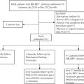 Efficacy and Safety of Focused Ultrasound Treatment for High-risk Human Papillomavirus Infection-related Cervical Intraepithelial Neoplasia Grade 2 in Nulligravidae Women: A Retrospective Study
 Assessing Intensive Care Unit Acquired Weakness: An Observational Study Using Quantitative Ultrasound Shear Wave Elastography of the Rectus Femoris and Vastus Intermedius
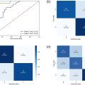 Development of a Deep Learning Model for Classification of Hepatic Steatosis from Clinical Standard Ultrasound
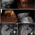 The Relationship Between Rim-like Enhancement on Pre-ablation Contrast-enhanced Ultrasound of Colorectal Liver Metastasis and Early Intrahepatic Progression After Thermal Ablation: A Preliminary Study
 Transcranial MRI-guided Histotripsy Targeting Using MR-thermometry and MR-ARFI
Stay updated, free articles. Join our Telegram channel

Full access? Get Clinical Tree


