Abstract
This case report presents a 38-year-old female with unilateral open-lip schizencephaly coexisting with septo-optic dysplasia. The patient lacked motor deficits, maintaining normal developmental milestones. schizencephaly is a rare cortical malformation characterized presence of abnormal cleft in the cerebral hemispheres of the brain. These clefts extend from the surface of the brain to the ventricles. Lissencephaly can be unilateral and bilateral and divided into open lips and closed lip. It is frequently associated with other anomaly such as septo-optic dysplasia, grey matter heterotopia, septum pellucidum, and dysgenesis of the corpus callosum. Schizencephaly has no known gender predilection, and estimated incidence of 1.5: 100,000 live birth. Radiological imaging is the cornerstone of diagnosis schizencephaly.
Introduction
Schizencephaly is an rare, congenital disorder affecting cerebral malformation characterized by clefts in the cerebral cortex, which extends from the surface of the ventricle through the white matter. It was first described in 1887, and 2 types exist: closed lip (Type I) and open lip (Type II). Type II is more common, especially in unilateral cases. Magnetic resonance was more sensitive than computed tomography in detection. Schizencephaly is associated in some cases with microcephaly, hydrocephalus, or other malformations such as septo-optic dysplasia and affects the posterior frontal and parietal lobes, with rare occurrences in the temporal or occipital lobe being disorder can be detected in vivo by ultrasonography [ ].
Scizencephaly can be associated with other malformations such as septo-optic dysplasia, grey matter heterotopia, septum pellucidum, and dysgenesis of the corpus callosum. Septo optic dysplasia characterized by underdevelopment of the unilateral or bilateral optic nerve and chiasma, along with the aplasia or hipoplasia of the septum pellucidum, and when associated with schizencephaly, it is referred to as septo-optic dysplasia plus [ ].
Case presentation
A 38 year old female patient was referred to the radiology department for an MRI Brain for proptosis since 5 year ago. She had history of blur vision since childhood. She did not have history of seizure. She had no history of limb weakness. There was no family history of neurological or psychiatric disorders, and there was no history of birth-related trauma or maternal exposure to medications. He was a nonsmoker, and nondrinker, and denied any recreational drug use.
On examination, the patient was oriented to time, place, and person, with normal cognitive function. Vital signs were within normal limits. There were no observed abnormalities in growth velocity or pubertal maturation. She had 2 children. Motor examination revealed was normal. Sensory examination was normal. Blood and laboratory test within normal range.
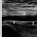
The Brain MRI revealed connection between parietal lobes right lateral ventricle via thin tract with hydrocephalus non communicans. Hypoplasia of optic nerves, absence of the septum pellucidum, thinning of corpus callosum body which features of septo optic dysplasia. The scan also indicated atrophy of right M. rectus medialis also increase size of long axis bulbus oculi bilateral. A diagnosis of unilateral schizencephaly open lip with septo optic dysplasia was made ( Figs. 1-5 ).

Related posts:
 A rare and life-threatening case of spontaneous hemopneumothorax presenting with severe hemodynamic instability
 A very rare case of ileocolic and appendiceal intussusception with acute appendicitis
 Hepatic and extra-hepatic hydatid cysts: A case series of radiological and clinical insights
 Mid-term follow-up of a fractured flow diverter in the internal carotid artery
 An atypical multiple autoimmune syndrome: A case report including myocarditis
 Successful ilio-femoral-popliteal venous aspiration thrombectomy using a 12 French system in a pediatric patient
Stay updated, free articles. Join our Telegram channel

Full access? Get Clinical Tree


