Unilateral/Bilateral Hand Motion
Lubdha M. Shah, MD
Key Facts
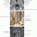
Imaging Anatomy
Unilateral sequential finger tapping results in robust activity within cortex surrounding superior lateral central sulcus in expected somatotopic location for finger/hand
Center of “hand knob” in precentral gyrus has been identified as landmark for hand area of primary motor cortex
Supplementary motor area and premotor activation may also be seen
Design
4-minute block design with visual stimulus presentation and “stop”/“go” commands
Different variations of paradigm
Repetitive finger tapping or hand squeezing results in robust premotor and primary sensorimotor activation
Tapping of thumb to each finger in sequential manner at self-paced rate on “go” command until “stop” command
Applications
Presurgical motor mapping for lesions in sensorimotor cortex
Patients with neurological cortical or spinal deficits may not be able to carry out finger-tapping tasks
Passive sensory stimulus may be used in subjects unable to perform hand movement
Subjects with abnormal evoked cerebral blood oxygenation changes due to arteriovenous malformation, stroke, or tumor may have false-negative activations (i.e., marked reductions of activation volumes) in BOLD imaging
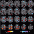
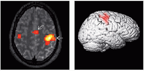 (Left) Axial fMRI in a neurologic format shows activation in the right sensorimotor region  due to a left-thumb flexion task. There is also activation in the supplementary motor area (SMA) due to a left-thumb flexion task. There is also activation in the supplementary motor area (SMA)  . Unilateral sequential finger tapping results in robust activity within the cortex surrounding the superior lateral central sulcus in the expected somatotopic location for the finger/hand. (Right) 3D surface-rendered image shows overlay of activation in the right sensorimotor region . Unilateral sequential finger tapping results in robust activity within the cortex surrounding the superior lateral central sulcus in the expected somatotopic location for the finger/hand. (Right) 3D surface-rendered image shows overlay of activation in the right sensorimotor region  due to the left-thumb flexion task. due to the left-thumb flexion task. |
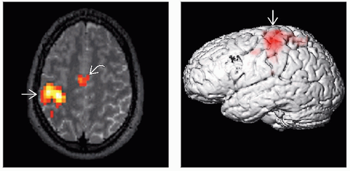 (Left) Axial fMRI in a neurologic format shows activation in the left sensorimotor region
 due to a right-thumb flexion task. There is also activation in the SMA due to a right-thumb flexion task. There is also activation in the SMA  . (Right) 3D surface-rendered image shows an overlay of activation in the left sensorimotor region . (Right) 3D surface-rendered image shows an overlay of activation in the left sensorimotor region  due to the right-thumb flexion task. Many studies have found high correlations between intraoperative cortical stimulation mapping with preoperative motor fMRI. due to the right-thumb flexion task. Many studies have found high correlations between intraoperative cortical stimulation mapping with preoperative motor fMRI.Related posts:Stay updated, free articles. Join our Telegram channel
Full access? Get Clinical Tree
 Get Clinical Tree app for offline access

|