Abstract
Spinal epidural abscess (SEA) is an uncommon infection typically caused by Staphylococcus aureus, predominantly affecting older adults with risk factors such as advanced age, comorbidities, and prior surgical interventions. This report details a case involving a 14-year-old male presenting with acute lumbar pain, lower limb weakness, urinary retention, and skin pustules. The patient had no history of immunologic diseases, diabetes, or drug use. Contrast-enhanced magnetic resonance imaging (MRI) with gadolinium demonstrated a peripheral fluid signal surrounding the thoracic spinal cord, consistent with an epidural abscess from T9-T12 to L1, causing spinal cord compression. Cultures from the abscess and skin pustules identified Staphylococcus epidermidis (S. epidermidis). The patient underwent surgical drainage and remained stable postoperatively. This case highlights an unusual SEA presentation in an adolescent without typical risk factors, emphasizing the importance of considering atypical pathogens and further investigation into infection sources.
Introduction
Spinal abscesses are categorized based on infection localization into 3 categories: epidural, subdural, and intramedullary [ ]. Spinal epidural abscess (SEA) is an uncommon infection characterized by the accumulation of purulent material within the space between the dura and vertebral periosteum [ ]. The most common microbial cause of SEA is Staphylococcus aureus, accounting for approximately two-thirds of cases. Other causative microbes involve coagulase-negative Staphylococcus (such as Staphylococcus epidermidis), Streptococcus, gram-negative bacilli (notably Pseudomonas aeruginosa and E. coli), and less frequently, anaerobic bacteria, fungi, mycobacteria, and parasites [ ]. Advanced age, comorbidities, and surgical procedures are common risk factors [ ]. SEA typically occur at a mean age of 50 years, mainly in those aged 30-70, with rare occurrences in children [ ]. Clinical presentation of neck or back pain, fever, potential neurological deficits, and magnetic resonance imaging (MRI) with gadolinium contrast are the diagnostic standards [ ]. SEA can result in spinal cord ischemia due to compression, leading to potentially life-threatening consequences [ ]. Therefore, prompt surgical drainage and administration of antibiotics are imperative in managing this critical medical condition [ ]. This paper describes a patient’s case who presented with acute back pain and neurological symptoms. Despite the absence of apparent risk factors, a thoracic-lumbar SEA due to Staphylococcus epidermidis (S. epidermidis) was diagnosed. The unique presentation of S. epidermidis without typical infection routes raises questions about the source of infection.
Case presentation
Chief complaints
A 14-year-old male patient presented to the neurological department with acute severe lumbar pain radiating to the lower limbs, where mild weakness progressed over 1 week to significant motor impairment.
Medical history and physical examinations
The patient has no history of immunologic diseases, diabetes, or medication use. Clinical examination showed 3/5 motor weakness in the lower limbs, 4/5 dorsal plantar flexion of the foot, urinary retention, diffuse skin pustules on the back of the thigh, shoulder, and humerus, and a little on the back.
Laboratory examinations
These pustules were cultured, revealing S. epidermidis as the causative pathogen. Laboratory test results revealed a marked increase in white blood cell (WBC; 15 × 109/l), erythrocyte sedimentation rate (ESR; 78.00 mm/h), and C-reactive protein (CRP; 123.96 mg/l) levels, suggesting a severe infection, with liver and kidney function tests normal and negative results for both the Vidal and Wright tests.
Imaging examinations and differential diagnosis
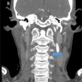
Based upon the neurologic symptomatology and blood test findings, the differential diagnosis was a compressive lesion in the lower dorsal spinal cord such as a tumor, disc, hemorrhage, or abscess. Contrast-enhanced MRI with gadolinium demonstrated a peripheral fluid signal surrounding the thoracic spinal cord, consistent with an epidural abscess from T9-T12 to L1, causing spinal cord compression ( Fig. 1 ). The recommendation for surgical excision was made, with the patient’s consent obtained and medical consultations prior to the procedure indicating no objections.

Surgical management and final diagnosis
Prophylactic antibiotics prior to surgery were made with intravenous vancomycin 1 g bi-daily and ceftriaxone 1 g bi-daily. The surgery was done by a specialist in neurosurgery, which involved T9-T11 laminectomy, canal dilatation, and SEA drainage via a posterior approach ( Fig. 2 ). Purulent fluid samples were collected and sent for Gram staining and bacteriological culture analysis. Irrigation and evacuation were performed multiple times until no abscesses or purulent material remained. The incision was repeatedly and carefully irrigated with iodine and saline mixed with vancomycin before closure, with the negative pressure drainage vessel remaining in situ. After surgery, cultures of the patient’s blood and drained pus revealed S. epidermidis, consistent with the pathogens detected in skin pustule tests and suggesting these lesions as the likely source of infection. Consequently, based on the final microbiological sensitivity report, the patient was prescribed oral cefixime 400 mg daily and oral amoxicillin 1 g bi-daily for 2 weeks.

Related posts:
 A rare and life-threatening case of spontaneous hemopneumothorax presenting with severe hemodynamic instability
 A very rare case of ileocolic and appendiceal intussusception with acute appendicitis
 Hepatic and extra-hepatic hydatid cysts: A case series of radiological and clinical insights
 Mid-term follow-up of a fractured flow diverter in the internal carotid artery
 An atypical multiple autoimmune syndrome: A case report including myocarditis
 Persistence of the hypoglossal artery: A rare case report
Stay updated, free articles. Join our Telegram channel

Full access? Get Clinical Tree


