Abstract
Carcinomas often metastasize to distant organs by detaching from the primary tumor and entering the bloodstream. These circulating tumor cells are transported to sites such as lung capillaries and subsequently disseminated through systemic circulation. However, hepatocellular carcinoma (HCC) rarely metastasizes to distant organs, even in advanced stages. Instead, HCC frequently leads to intravascular and intrahepatic parenchymal metastases. Portal vein thrombosis (PVT), present in 10%-40% of HCC patients at diagnosis and 35%-44% at death or liver transplant, is a significant prognostic marker, associated with worse outcomes and reduced survival. Patients with PVT demonstrate markedly shorter overall survival compared to those without PVT, with main portal vein thrombosis indicating the poorest prognosis. Here, we report an unusual case of an enormous portal metastasis from liver HCC, characterized by a “vessel-inside-vessel” appearance.
Case report
A 63-year-old woman with a medical history of familial hyperlipidemia underwent a routine health examination, including liver ultrasonography (US), for hyperlipidemia monitoring. This revealed multiple hypodense hepatic lesions. Physical examination and laboratory investigations were unremarkable, prompting further imaging with an abdominal computed tomography (CT) scan.
Contrast-enhanced abdominal CT was performed during the arterial, portal, and delayed phases. Multiple hypodense hepatic lesions with vivid enhancement in the arterial phase and washout in the delayed phase were observed, alongside areas of central necrosis. The enhancement pattern was consistent with hepatocellular carcinoma.
Additionally, a voluminous mass was identified within the splenic vein ( Figs. 1 and 2 ), extending into the portal trunk and causing extensive collateral venous circulation in the gastro-splenic region ( Fig. 3 ). The mass demonstrated vivid arterial-phase enhancement, angiogenesis within the thrombus (“vessel-inside-vessel” appearance), and late-phase washout, mirroring the enhancement pattern of the hepatic lesions.


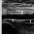
Ultrasonography revealed a hypoechoic mass within the main portal trunk and splenic vein ( Fig. 4 ). Color Doppler imaging showed hypervascularity ( Fig. 5 ), with arterial and venous flow confirmed on spectral Doppler. A percutaneous ultrasound-guided biopsy was performed, and histopathological examination confirmed poorly differentiated hepatocellular carcinoma.

Related posts:
 A rare and life-threatening case of spontaneous hemopneumothorax presenting with severe hemodynamic instability
 A very rare case of ileocolic and appendiceal intussusception with acute appendicitis
 Hepatic and extra-hepatic hydatid cysts: A case series of radiological and clinical insights
 Mid-term follow-up of a fractured flow diverter in the internal carotid artery
 An atypical multiple autoimmune syndrome: A case report including myocarditis
 Successful ilio-femoral-popliteal venous aspiration thrombectomy using a 12 French system in a pediatric patient
Stay updated, free articles. Join our Telegram channel

Full access? Get Clinical Tree


