Vascular Closure Devices


Vascular Closure Devices


Evan Lehrman

Joshua L. Weintraub


Safe and effective arterial access closure and hemostasis continue to present a challenge for interventional radiologists. Arterial access is required for a wide range of procedures ranging from interventional oncology and peripheral arterial disease to aneurysm repair and trauma. Arterial access sheaths range from 4 Fr. for diagnostic angiography to 12 to 25 Fr. for endograft delivery. Although manual compression (MC) has been the gold standard for achieving hemostasis for smaller arteriotomies since the inception of interventional radiology, vascular closure devices (VCDs) were introduced in the early 1990s, and a large body of evidence and experience has accumulated to support their use. Advantages and disadvantages exist for both methods of arteriotomy closure (Table 3.1).


Table 3.1 Advantages and Disadvantages of Manual Compression and Vascular Closure Devices


Method


Advantages


Disadvantages


Manual compression


-Gold standard


-Limited learning curve


-No device cost


-Patient discomfort


-Delayed time to hemostasis


-Delayed time to ambulation


-Anticoagulation must be reversed (ACT <180)


-Decreased workflow


Vascular closure devices


-Immediate hemostasis


-Hemostasis possible with anticoagulation


-Decreased time to ambulation


-Improved patient comfort


-Improved workflow may offset device costs


-Device cost


-Learning curve for each device


-Immediate repuncture not recommended for all devices


-Potential for severe complications


Types of Devices

Several categories of VCD are currently manufactured, each with their own mechanism of action, applications, and learning curves.

1. Active approximators (Table 3.2)

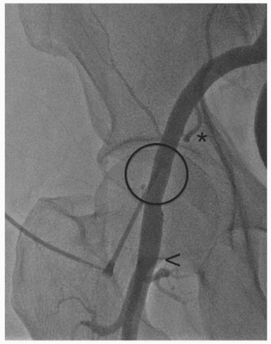
These devices mechanically close the arteriotomy as material is fixed to the vessel wall. With Angio-Seal, the arteriotomy is sandwiched between an intravascular footplate and collagen plug. With suture- and clip-mediated devices, the arteriotomy itself is cinched closed. Active approximators are considered the most secure form of VCD but carry the highest risk of serious complication.

2. Passive approximators (Table 3.3)

These devices deploy a sealant, gel, or plug at the extravascular aspect of the arteriotomy to achieve hemostasis. Adjunctive MC is often held for 1 to 3 minutes. These devices have the highest risk of incomplete hemostasis.

3. Nondepositional hemostasis aids (Table 3.4)

This is a heterogeneous group of devices that do not leave any material behind at the end of the procedure.

4. Hemostatic patches (Table 3.5)

These patches are placed externally over the dermatotomy prior to MC. Substances interact with blood products to aid in hemostasis.

5. Mechanical compression aids (Table 3.6)

These devices allow MC to be performed by the device rather than an operator.


Table 3.2 Active Approximators


Device


Manufacturer


Mechanism


Sheath Size (Fr.)


Comments


StarClose SE


Abbott Vascular


Clip


5, 6


Extravascular clip cinches arteriotomy closed


Angio-Seal


St. Jude Medical


Footplate/plug


6, 8


Arteriotomy sandwiched between footplate and collagen plug


-VIP


-Evolution


Perclose


Abbott Vascular


Suture


5-8


Braided polyester or monofilament sutures


-ProGlide


-A-T


Prostar XL


Abbott Vascular


Suture


8.5-10


Braided polyester suture, used for larger sheaths


FemoSeal


St. Jude Medical


Intra- and extravascular discs


7


Biodegradable polymer discs


Table 3.3 Passive Approximators


Device


Manufacturer


Mechanism


Sheath Size (Fr.)


Comments


Mynx


Access Closure


Sealant


5-7


Polyethylene glycol deployed while intravascular balloon creates temporary hemostasis


-Grip


-Ace


FISH


Morris Innovative


Sealant


5-8


Extracellular matrix closure patch premounted on procedural sheath


EXOSEAL


Cordis


Sealant


5-8


Polyglycolic acid deployed through existing sheath


FastSeal


Vascular Closure Systems


Bioabsorbable plug


6, 8


Plug deployed through existing sheath. Safety tether allows removal if malpositioned.


VASCADE


Cardiva


Collagen plug


5-7


Collagen plug deployed while intravascular mesh disc creates temporary hemostasis


Only gold members can continue reading. Log In or Register to continue

Stay updated, free articles. Join our Telegram channel

Jun 17, 2016 | Posted by in INTERVENTIONAL RADIOLOGY | Comments Off on Vascular Closure Devices

Full access? Get Clinical Tree

Get Clinical Tree app for offline access

Table 3.4 Nondepositional Hemostasis Aids