IMAGING ANATOMY
Overview
- •
Vertebral artery (VA): 4 segments
- ○
V1 segment (extraosseous segment)
- –
Arises from 1st part of subclavian artery
- –
Courses posterosuperiorly to enter C6 transverse foramen
- –
Branches: Segmental cervical muscular, spinal branches
- –
- ○
V2 segment (foraminal segment)
- –
Ascends through C6-C3 transverse foramina
- –
Turns superolaterally through inverted L-shaped transverse foramen of axis (C2)
- –
Courses short distance superiorly through C1 transverse foramen
- –
Branches: Anterior meningeal artery, unnamed muscular/spinal branches
- –
- ○
V3 segment (extraspinal segment)
- –
Exits top of atlas (C1) transverse foramen
- –
Lies on top of C1 ring, curving posteromedially around atlantooccipital joint
- –
As it passes around back of atlantooccipital joint, turns sharply anterosuperiorly to pierce dura at foramen magnum
- –
Branches: Posterior meningeal artery
- –
- ○
V4 segment (intradural/intracranial segment)
- –
After VA enters skull through foramen magnum, courses superomedially behind clivus
- –
Unites with contralateral VA at or near pontomedullary junction to form basilar artery (BA)
- –
Branches: Anterior, posterior spinal arteries (ASA, PSA), perforating branches to medulla, posterior inferior cerebellar artery (PICA)
- –
Arises from distal VA, curves around/over tonsil, gives off perforating medullary, choroid, tonsillar, cerebellar branches
- –
- ○
- •
BA
- ○
Courses superiorly in prepontine cistern (in front of pons, behind clivus)
- ○
Bifurcates into its terminal branches, posterior cerebral arteries (PCAs), in interpeduncular or suprasellar cistern at or slightly above dorsum sellae
- ○
Branches: Pontine, midbrain perforating branches (numerous), anterior ICA (AICA), superior cerebellar arteries (SCAs), PCAs (terminal branches)
- ○
Vascular Territory
- •
VA
- ○
ASA: Upper cervical spinal cord, inferior medulla
- ○
PSA: Dorsal spinal cord to conus medullaris
- ○
Penetrating branches: Olives, inferior cerebellar peduncle, part of medulla
- ○
PICA: Lateral medulla, choroid plexus of 4th ventricle, tonsil, inferior vermis/cerebellum
- ○
- •
BA
- ○
Pontine perforating branches: Central medulla, pons, midbrain
- ○
AICA: Internal auditory canal, CNVII, and CNVIII, anterolateral cerebellum
- ○
SCA: Superior vermis, superior cerebellar peduncle, dentate nucleus, brachium pontis, superomedial surface of cerebellum, upper vermis
- ○
Normal Variants, Anomalies
- •
Normal variants
- ○
VA: Variation in size from right to left, dominance common; origin from aortic arch in 5%
- ○
- •
Anomalies
- ○
VA/BA may be fenestrated or duplicated (may have increased prevalence of aneurysms)
- ○
Embryonic carotid-basilar anastomoses (e.g., persistent trigeminal artery)
- ○
ANATOMY IMAGING ISSUES
Imaging Recommendations
- •
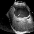
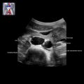
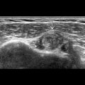
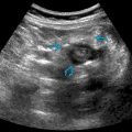
V1 and V2 segments are amenable to US examination
- •
Examination usually starts in V2 segment and proceeds downward to V1 segment, then to its origin
- •
Examination of V2 segment
- ○
Transducer oriented longitudinally in midcervical region between trachea and sternocleidomastoid muscle
- ○
Angle transducer laterally from common carotid artery (CCA) and locate V2 segment posterior to acoustic shadowing of transverse processes
- ○
- •
Examination of V1 segment
- ○
Trace caudally from V2 to its origin
- ○
Left VA more difficult to visualize than right VA
- ○
Do not confuse with vertebral vein lying adjacent to VA, which can appear pulsatile
- –
Color flow imaging helps to differentiate
- –
- ○
- •
Normal waveform of VA on spectral Doppler analysis
- ○
Low-resistance flow
- ○
Similar to that of CCA but with lower amplitude
- ○
PSV: 59 ± 17 cm/s; EVD: 19 ± 8 cm/s
- ○
Flow velocity asymmetry is common and related to caliber of VA
- ○
Imaging Pitfalls
- •
VA distal to V2 cannot be properly assessed by US
- ○
Abnormalities in spectral Doppler waveform of VA at V1/V2 segment provide clue for disease beyond V2
- ○
EMBRYOLOGY
Embryologic Events
- •
Plexiform longitudinal anastomoses between cervical intersegmental arteries → VA precursors
- •
Paired plexiform dorsal longitudinal neural arteries (LNAs) develop, form precursors of BA
- •
Transient anastomoses between dorsal LNAs develop, and ICAs appear (primitive trigeminal/hypoglossal arteries, etc.)
- •
Definitive VAs arise from 7th cervical intersegmental arteries, anastomose with LNAs
- •
LNAs fuse as temporary connections with ICAs regress → definitive BA, vertebrobasilar circulation formed
GRAPHICS AND VOLUME-RENDERED CTA