The Artery of the Filum Terminale
42.1 Case Description
42.1.1 Clinical Presentation
A 62-year-old man presented with a 6-month history of progressive bilateral lower extremity paresthesias and weakness. Three months before admission, bladder incontinence developed, and 1 month later, the patient became impotent.
42.1.2 Radiological Studies
See ▶ Fig. 42.1, ▶ Fig. 42.2.

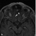
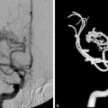
Fig. 42.1 Sagittal T2-weighted (a) MRI demonstrated venous congestion with vasogenic edema of the spinal cord and swelling from the lower thoracic levels to the conus medullaris, along with flow voids suggestive of enlarged perimedullary veins. Contrast-enhanced T1-weighted (b,c) sequences revealed a moderate degree of diffuse enhancement of the lower thoracic spinal cord, indicative of chronic venous ischemia. Contrast-enhanced MRA (d,e,f) of the spine showed a dilated midline vessel, likely the ASA, down to the filum terminale. The patient underwent conventional angiography for further evaluation. Case continued in ▶ Fig. 42.2.

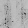
Fig. 42.2 Right T8 segmental artery angiogram (a–d) demonstrates an abnormally enlarged and tortuous anterior spinal artery leading to an arteriovenous fistula that is located well below the cone at the level of the filum terminale at S2–S3. The single fistulous communication was noted to occur between the artery and the vein of the filum terminale with venous outflow in a cranial direction along the filum terminale, draining cranially into the perimedullary veins.
42.1.3 Diagnosis
Spinal arteriovenous fistula of the filum terminale.
42.2 Embryology and Anatomy
The primordia of the filum terminale forms during secondary neurulation. This process occurs after closure of the caudal neuropore. Degeneration, regression, and retrogressive differentiations of the secondary neural tube then lead to the development of the filum terminale, which remains as a fibrous structure connecting the conus medullaris to the coccyx. The filum terminale has two components. The filum interna is an extension of the primary pia mater that connects the conus medullaris to the dura mater of the terminal thecal sac. The filum externa extends from the termination of the thecal sac to the coccyx.
Classically, the arterial supply to the filum terminale was described as singular, fed by the artery of the filum terminale. This artery is the caudal extension of the anterior spinal artery, which arises at the conus medullaris. Most commonly, the anterior spinal artery bifurcates at the conus medullaris and forms the anastomotic arcade, and the artery of the filum arises from one of the branches of the bifurcation. Anatomic variants have been described, including a trifurcation of the anterior spinal artery, with one of the branches continuing caudally as the artery of the filum.
Related posts:


Stay updated, free articles. Join our Telegram channel

Full access? Get Clinical Tree


