Basics of Imaging: Radiography, CT, and MR
QUESTIONS
1 What is the purpose of double-inversion recovery in black blood imaging?
A. To improve blood pool signal
B. To suppress fat
C. To suppress blood flow
D. To improve temporal resolution
View Answer
1 Answer C. Double-inversion recovery sequence in black blood cardiac imaging is designed to suppress the signal from blood flow.
Reference: Ginat DT, Fong MW, Tuttle DJ, et al. Cardiac imaging: part 1, MR pulse sequences, imaging planes, and basic anatomy. AJR Am J Roentgenol 2011;197(4):808-815. doi: 10.2214/AJR.10.7231.
2 With conventional filtered-back projection (FBP), what is the relationship of tube current to noise?
A. Directly proportional
B. Inversely proportional
C. No direct relationship
D. Exponentially proportional
View Answer
2 Answer B. With filtered-back projection, tube current is inversely proportional to noise. That is, increasing the mA by factor of 4 will yield half the noise (1/square root of 4). Tube current determines the number of photons generated and noise.
Reference: Litmanovich DE, Tack DM, Shahrzad M, et al. Dose reduction in cardiothoracic CT: review of currently available methods. Radiographics 2014;34(6):1469-1489. doi: 10.1148/rg.346140084.
3 A patient is coming back for a follow-up CT. You looked at a prior CT, and it was very noisy. What parameter can you change on the follow-up CT to reduce the noise by a factor of 2 (assuming filtered-back projection was used)?
A. Increase the effective mAs by a factor of 2.
B. Increase the effective mAs by a factor of 4.
C. Decrease the kVp by 40%.
D. Decrease the kVp by 20%.
View Answer
3 Answer B. With filtered-back projection, tube current is inversely proportional to noise. That is, increasing the mA by factor of 4 will yield half the noise (1/square root of 4). Relationship of kVp to noise is complex, but in general, decreasing the kVp will increase the noise if other factors are held constant.
Reference: Litmanovich DE, Tack DM, Shahrzad M, et al. Dose reduction in cardiothoracic CT: review of currently available methods. Radiographics 2014;34(6):1469-1489. doi: 10.1148/rg.346140084.
4 Assuming a rotation time of 0.3 seconds and mA of 700, what is the effective tube current-time product if the pitch is 0.2?
A. 2,100 mA
B. 1,050 mA
C. 210 mA
D. 42 mA
View Answer
4 Answer B. Effective tube current-time product is obtained by multiplying the rotation time by the mA and then dividing by the pitch. So in this case 0.3 × 700 = 210 mA, which is then divided by 0.2, giving 1,050 mA.
Reference: Litmanovich DE, Tack DM, Shahrzad M, et al. Dose reduction in cardiothoracic CT: review of currently available methods. Radiographics 2014;34(6):1469-1489. doi: 10.1148/rg.346140084.
5 In filter-back projection, changing the type of reconstruction algorithm/kernel can affect the spatial resolution and what else?
A. Radiation dose
B. Noise of image
C. Temporal resolution
View Answer
5 Answer B. Reconstruction algorithm/kernel does not affect radiation dose since it is applied after the study is already obtained. It can affect spatial resolution and noise depending on which algorithm/kernel is used.
Reference: Litmanovich DE, Tack DM, Shahrzad M, et al. Dose reduction in cardiothoracic CT: review of currently available methods. Radiographics 2014;34(6):1469-1489. doi: 10.1148/rg.346140084.
6 How is dose length product (DLP) related to scan length?
A. It is not related.
B. It is directly proportional.
C. It is inversely proportional.
View Answer
6 Answer B. DLP is obtained by multiplying the CTDIvol by the scan length; therefore, it is directly proportional.
Reference: Litmanovich DE, Tack DM, Shahrzad M, et al. Dose reduction in cardiothoracic CT: review of currently available methods. Radiographics 2014;34(6):1469-1489. doi: 10.1148/rg.346140084.
7 How does one calculate an estimated effective dose in millisieverts?
A. Multiply the dose length product by a conversion factor.
B. Divide the dose length product by a conversion factor.
C. Multiply the CT volume dose index by a conversion factor.
D. Divide the CT volume dose index by a conversion factor.
View Answer
7 Answer A. Effective dose gives a general population risk rather than patient-specific risk. It is obtained by multiplying the DLP by a conversion factor (f). The conversion factor is obtained by Monte Carlo simulation, and the best estimates (f) factor should be size specific.
Reference: Litmanovich DE, Tack DM, Shahrzad M, et al. Dose reduction in cardiothoracic CT: review of currently available methods. Radiographics 2014;34(6):1469-1489. doi: 10.1148/rg.346140084.
8 In a patient with contraindication to beta-blockers, which medication can be given to slow the heart rate?
A. Atenolol
B. Nitroglycerin
C. Verapamil
D. Sildenafil
View Answer
8 Answer C. In patients with contraindication to beta-blocker (such as second-degree heart block, severe asthma, decompensated heart failure), a calcium channel blocker can be used. Verapamil is a calcium blocker agent. Atenolol is a beta-blocker so it should not be used if there is contraindication to beta-blocker. Nitroglycerin is used for vasodilatation of the coronaries and will not slow the heart rate. Sildenafil (Viagra) should not be used concurrently with nitroglycerin as it could cause severe hypotension.
Reference: Taylor CM, Blum A, Abbara S. Patient preparation and scanning techniques. Radiol Clin North Am 2010;48(4):675-686. doi: 10.1016/j.rcl.2010.04.011.
9 The image below is from a phase-contrast image in a patient with suspected pulmonic stenosis. Which of the following statements is most accurate about the image?
|
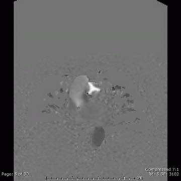
A. The velocity-encoding gradient was set too low.
B. The image shows no net phase shift of the blood.
C. Bipolar gradients were applied to obtain the image.
D. There is stenosis of flow across the valve.
View Answer
9 Answer C. Phase-contrast images are used to measure blood flow and velocity. In cardiac imaging, they are most commonly used to evaluate the peak velocity in cases of valve stenosis and the regurgitant fraction in cases of valve insufficiency. A bipolar gradient is applied, and results in stationary objects experiencing no net phase shift while moving objects will experience a phase shift proportional to their velocity, which yields signal. If the velocity-encoding gradient is set too high or low, aliasing will occur (which is not on the image below).
|

Reference: Lotz J, et al. Cardiovascular flow measurement “with phase-contrast MR imaging: basic facts and implementation. Radiographics 2002;22(3):651-671.
10 Your department needs a new CT scanner in the emergency department and wants to offer cardiac CTA. A vendor says the single-source scanner has a temporal resolution of 200 msec when using a single-segment reconstruction. If this statement is true, what must the rotation speed of the scanner be?
A. 200 msec
B. 300 msec
C. 400 msec
D. 500 msec
View Answer
10 Answer C. With a single-source CT scanner, the image can be generated once 180 degrees of data have been acquired. The temporal resolution is calculated by dividing the rotation speed of the scanner by 2 (Temporal resolution = Rotation speed/2). In our example, 200 msec = Rotation speed/2; 400 msec = Rotation speed.
Reference: Lin E, Alessio A. What are the basic concepts of temporal, contrast, and spatial resolution in cardiac CT. Cardiovasc Comput Tomogr 2009;3(6):403-408.
11 A 55-year-old male with a history of atypical chest pain undergoes a retrospective cardiac CTA at your institution on a 64-slice scanner. As you inject contrast, the heart rate increases to 85 beats per minute during the entire scan acquisition. Your technologist reconstructs the data, and you are still able to interpret the exam. What strategy did your technologist employ?
A. Use a sharp kernel/filter.
B. Multi-segment reconstruction.
C. Increased the pitch during the exam.
D. Use tube modulation.
View Answer
11 Answer B. Patients with an elevated heart rate who undergo retrospective cardiac CTA can have the data analyzed using multisegment reconstruction techniques. When using multisegment reconstruction, the image will be created using data from multiple heart beats. This will produce an image that potentially has better temporal resolution than single segment reconstruction. Multisegment reconstruction requires the study be acquired with a low pitch. The use of multiple heart beats makes this techniques susceptible to motion artifact and heart rate variability.
Reference: Mahesh M, Cody DD. Physics of cardiac imaging “with multiple-row detector CT. Radiographics 2007;27(5):1495-1509.
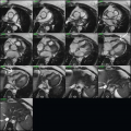
12 Your technologist completes a short-axis balanced steady-state free precession cardiac MRI (cMRI) sequence to calculate cardiac function. While scanning the apex of the heart, the technologist notices a mass in the liver with bright signal and asks you if this sequence can confidently characterize the lesion. Your response is which of the following?
A. Yes, since the sequence is only T2 weighted, the mass is a cyst.
B. Yes, it is a cyst since the sequence is not susceptible to calcification or metallic artifact.
C. No, the mass contains calcification, which accounts for its bright signal.
D. No, although the sequence has relative T2 weighting, it has both T2 and T1 properties.
View Answer
12 Answer D. The balanced steady-state free precession sequence is a gradient-echo sequence that is susceptible to metallic artifact and has weighted T2/T1 signal. While the sequence is relatively T2 weighted, it will also have T1 properties.
References: Chavan GB, Babyn PS, Jankharia BG, et al. Steady-state MR imaging sequences: physics, classification, and clinical applications. Radiographics 2008;28(4)1147-1160.
Bieri O, Scheffler K. Fundamentals of balanced steady state free precession MRI. J Magn Reson Imaging 2013;38:2-11. doi: 10.1002/jmri.24163.
13 A postprocessing technique that chooses the maximum voxel value in a defined thickness and uses it as the displayed value is called
A. Curved multiplanar reformatted image
B. Maximum-intensity projection image
C. Shaded surface display image
D. Volume-rendered image
View Answer
13 Answer B. The maximum-intensity projection image uses the maximum voxel value to create a displayed value. The technique is useful to evaluate vessels; however, if the vessel is densely calcified or if there is metallic material, this technique may obscure the vessel lumen.
Reference: Calhoun PS, Kuszyk BS, Heath DG, et al. Three-dimensional volume rendering of spiral CT data: theory and method. Radiographics 1999;19(3):745-764.
14 A patient undergoes a cardiac CTA. The patient has no coronary artery disease in the vessels; however, while postprocessing the data, your 3D tech makes a pseudolesion in the left anterior descending coronary artery. Which post processing technique did your 3D tech most likely used?
A. Curved planar reformat
B. Maximum-intensity projection
C. Minimal-intensity projection
D. Volume-rendered
View Answer
14 Answer A. The abnormality most likely occurred on the curved planar-reformatted image. This technique places the long axis of the vessel (i.e., coronary artery) on a single image, allowing it to be visualized along its entire course. It allows stenosis to be readily visualized; however, it is susceptible to pseudolesions from an inability to show the vessel along its true long axis. This can result from unsuccessful vessel extraction, or motion artifact.
Reference: Dalrymple NC, Prasad SR, Freckleton MW, et al. Introduction to the language of three-dimensional imaging with multidetector CT. Radiographics 2005;25(5):1409-1428.
15 A patient arrives for a cardiac MRI to evaluate the mitral valve and aortic valve. Your sequence has a TR of 5 msec and the views per segment is 20. What is the temporal resolution of your scan?
A. 4 msec
B. 15 msec
C. 25 msec
D. 100 msec
View Answer


15 Answer D. Temporal Resolution = TR × Views per segment Temporal resolution is determined by how quickly the image is obtained (like shutter speed in a camera). Better temporal resolution is required to visualize fast moving structures such as valve leaflets.
References: Lee VS. Cardiovascular MRI: Physical principles to practical protocols. Lippincott Williams & Wilkins, 2006:291.
Related posts:
Stay updated, free articles. Join our Telegram channel
Full access? Get Clinical Tree
