, Yves Etienne2 and Maurice Niddam3
(1)
Faculté de Médecine, Marseille, France
(2)
Unité de Médecine Légale, Hôpital de la Timone, Marseille, France
(3)
Unité SAMU 13, Centre 15, Hôpital de la Timone, Marseille, France
The cerebral amygdala is connected to the septum, the diencephalon and brainstem via two pathways which are a dorsal pathway, the stria terminalis, and a ventral pathway called the ventral amygdalofugal pathway1. These pathways include both centripetal fibres to the amygdala (amygdalopetal fibres) and centrifugal fibres (amygdalofugal fibres), arising from the amygdala’s neurons. Their point of origin can therefore be described either from the targets of the amygdala fibres or from the amygdala itself. It is this second option that we have chosen. However, an inter-amygdaloid pathway of great importance should be added, the anterior commissure, which is included in this chapter.
6.1 The Dorsal Connection Pathway or Stria Terminalis
The stria terminalis is the current name of the former pathway of Foville2 or “taenia of Tarin”3 and became over time the semi-circular tenia (tenia semi-circulaire) according to French authors. It has been studied in depth as early as 1895 by J Déjerine (1901) and in parallel by A Koelliker (1896). It is a band of white substance, which appears as a flattened, very thin, 2–3 mm-wide band, with a fairly homogeneous diameter before it divides. It is very fragile and therefore difficult to dissect. Its length (6.6 cm, on average) slightly varies from one individual to another. It connects the amygdala to the septal region and to the hypothalamus. It is formed of a pathway of long fibres among which several are myelinated and whose origins are at the level of the nuclei of the amygdala, such as has already been demonstrated in animals, by CA Fox in 1943, T Ban and F Omukai in 1959, E Hall in 1960 and WJH Nauta in 1961. According to WM Cowan et al. 1965, only cortical and medial nuclei appear to be involved in the stria terminalis. Our observations made with humans have shown that, in fact, all of the nuclei are involved (Fig. 6.1) with a predominance for the medial nucleus. Yet, the stria terminalis appears to laterally leave the amygdala, which is paradoxical at first glance, since it emerges from the lateral slope of the superior pole (where is located the medial nucleus!). Such as Ch Foix and J. Nicolesco (1925) had already noted, the partitions between the different nuclei of the amygdala or surrounding them, as well as the peripheral capsule from which depart these partitions, are white substance lamina, formed by the neurofibres of neurons forming the nuclei. The stria thus results from the assembly of all the axons of the amygdaloid neurons, which are joined by the axons of the neurons from the centres, which it connects to the amygdala. The stria terminalis then follows the trajectory of the concave edge of the caudate nucleus. It initially runs along the medial surface of the tail of this formation. It will firstly progress to the rear, pass in the depth of the endorhinal sulcus, at the level of the uncus of the hippocampus, to reach the roof of the temporal lorn of the lateral ventricle, parallel to the fimbria from which it is only separated by a few millimetres. At the junction of the body and head of the hippocampus, the tenia of the fimbria and that of the stria terminalis unite (H.M. Duvernoy, in 1988), thus forming a small crescent moon-shaped lamella: the velum terminale of Aeby4 (Fig. 6.2). The choroidal web of the temporal horn of the lateral ventricle is attached to the above tenia and leaves the head of the hippocampus free and therefore not covered by the choroid plexi, unlike the other parts (body and tail) of this formation. The stria terminalis penetrates the opto-striated groove (or thalamo-striate sulcus), at the junction of the posterior thalamic convexity and concavity of the caudate nucleus. It therefore passes in the embryological joining area between the caudate nucleus (forebrain) and the thalamus (diencephalon). The stria terminalis is located in this groove successively in front of the thalamo-striate superior vein, at the level of the body of the caudate nucleus and below this vein, at the head of this nucleus. It is recalled that the superior thalamo-striate vein (terminal vein) drains the veins of the septum pellucidum and of the caudate nucleus and joins the homolateral superior choroidal vein to form one of the two internal cerebral veins, which are the roots of the great cerebral vein. The stria terminalis and its satellite vein will jointly form the bend with an anterior concavity of the thalamo-striate groove and thus reach the floor level of the frontal horn of the lateral ventricle, under the head of the caudate nucleus. They are covered, at this level, by the lamina affixa (lamina cornea), a thin epithelial layer, on the medial border of which are attached the choroid plexi of the lateral ventricle. Thus, the stria terminalis, over its arch-shaped trajectory, forms a 3/4 circle whose diameter is of 2.2 cm on average. Over this trajectory, the stria terminalis not only develops some thin branches, which target the caudate nucleus and the thalamus, but also a significantly sized branch which penetrates the latter at the level of the anterior third of the dorsal surface (Fig. 6.3). According to our chosen description approach, we can say that these branches contain input fibres targeting the thalamus. However, the opposite is also true and we can also say that these branches contain fibres from the thalamus, which are going to merge with the stria terminalis.
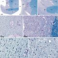
Fig. 6.1
The stria terminalis in the closest segment to amygdala (segment of origin or segment of ending, according to the considered fibres). (a, b) Two amygdalae (Amn) whose nuclei are dissected, with their own stria terminalis (strt). (c) Dissection and separation of the nuclei of an amygdala whose stria terminalis (strt) receives fibres’ fasciculi from each nucleus (NB: 10 nuclei have been highlighted). (d) Junction between the amygdala and the stria terminalis (Loyez staining for highlighting the myelin sheaths of nerve fibres). (e, f) Luxol-fast-blue-PAS stain (×100, ×200) reveals some scattered neurons between the fibres and the glial cells. The white arrows on (a) and (b) show two intercalated nuclei
Fig. 6.2
Medial aspect of the right cerebral hemisphere, showing the rostral division of the stria terminalis. Amn amygdaloid nuclear complex, 2 baso-lateral, 3 baso-medial, 4 cortical, CiG cingulate gyrus, cnb caudate nucleus body, cnh caudate nucleus head, cnt caudate nucleus tail, DG dentate gyrus, faG fasciolaris gyrus, Fc column of fornix, fc fasciola cinerea, Fcr crus of the fornix, Fi fimbria, gCC genu of the corpus callosum, GR gyrus rectus, sCG subcallosum gyrus, ssplG subsplenial gyrus, Hy hypothalamus, ICa internal capsule, iThad interthalamic adhesion, Lvac lateral ventricule anterior cornu, MB mamillary body, mlst medial longitudinal stria, pHG parahippocampal gyrus, rCC rostrum of the corpus callosum, splCC splenium of the corpus callosum, ssplG subsplenial gyrus, strt stria terminalis, a ending fasciculus (supra-commissural component) destinated to the paraterminalis gyrus and to the accumbens and septal nuclei, b other ending fasciculus (hypothalamic component) destinated to the anterior commissure, the fornix and the hypothalamus), tCC truncus of the corpus callosum, Th thalamus, Tpo temporal pole, Un a uncal apex, II optic nerve, Red arrow it shows the commissural component of strt entering the Gratiolet’s canal (the anterior commissure has been removed in order to see the fibres of this commissural component). Little black circles hypothalamic sulcus, white asterisk velum terminale of Aeby. The posterior portion of the diencephalon has been widely removed in order to show very well the stria terminalis. It is the same for the fornix’s body. The rostral part of the septum pellucidum is tracted up by a little retractor. A forceps pulls lightly the hypothalamus’s floor
Fig. 6.3
The thalamic ramus of the stria terminalis, highlighted on two dissections of the medial face of the left hemisphere (the ramus is visible on the centre of each red circle). (a) Thalamus in position; (b) thalamus resected. ACo anterior commissure, Cif cingulate fasciculus, CiG cingulate gyrus, cnb caudate nucleus, body, cnh caudate nucleus, head, ColS collateral sulcus, Fc column of fornix, gCC genu of the corpus callosum, Hy hypothalamus, ICa internal capsule, Ithad interthalamic adhesion, lOTG lateral occipito-temporal gyrus, lt lamina terminalis, LVac lateral ventricle anterior cornu, phG parahippocampal gyrus, pul pulvinar, rCC rostrum of the corpus callosum, sCG subcallosum gyrus, splCC splenium of the corpus callosum, stm stria medullaris, strt stria terminalis, tCC truncus of the corpus callosum, Th thalamus, Tpo temporal pôle, Un uncus
When the stria reaches the vicinity of the anterior commissure, it will be divided into three main pathways, which can be accessed for dissection (Fig. 6.2): the supra-commissural pathway (or precommissural component), the commissural pathway (commissural component) and the sub-commissural pathway (or retro-commissural component). These three pathways will develop as follows:
Get Clinical Tree app for offline access

The supra-commissural pathway passes above and in front of the anterior commissure and joins the septum to terminate in the septal nuclei (lateral nucleus and medial nucleus) (JS De Olmos 2004), and in the large nucleus accumbens septi, a key component of the pleasure and reward circuit. A part of the pathway reaches the cortex and terminates in the anterior perforated substance in the olfactory tubercle (G Paturet 1964) and in the paraterminal gyrus.
The fibres of the commissural pathway (commissural component) (Fig. 6.4) join the anterior commissure, thus forming communication pathways between the two brain hemispheres including the two amygdalae.
Fig. 6.4
The commissural component of the stria terminalis on two medial views (a) and (b) of the dissected right hemisphere. Amn amygdaloid nuclear complex, 2 baso-lateral nucleus, 3 baso-medial nucleus, 4 cortical nucleus, 5 central nucleus, 6 medial nucleus, bmFa baso-medial frontal artery, cF column of fornix, chci chiasmatic cistern, Cif cingulate fasciculus, CiG cingulate fasciculus, cnb caudate nucleus body, cnh caudate nucleus head, cnt caudate nucleus tail, Fb fornix body, fc fasciola cinerea, Fcr crus of the fornix, Fi fimbria, gCC genu of the corpus callosum, Hb hippocampal body, Hh hippocampal head, Ht hippocampal tail, Hy hypothalamus, ICa internal capsule, iThad interthalamic adhesion, llst lateral longitudinal stria, lt lamina terminalis, LVac lateral ventricle, anterior cornu, LVb lateral ventricle body, LVic lateral ventricle inferior cornu, MB mamillary body, mD margo denticulatus, Mes mesencephalon, och optic chiasma, ot optic tract, perica pericallosal artery, r’ optic recess, rCC rostrum of the corpus callosum, sCG subcallosum gyrus, Sepe septum pellucidum, strt stria terminalis, tCC truncus of the corpus callosum, Th thalamus, Tpo temporal pole, Un a apex of the uncus, UnF uncinate fasciculus, *Fb The asterisk shows the section’s level of the fornix body, II optic nerve, a1 anterior cerebral artery, black circles hypothalamic sulcus, red curved arrow anterior commissure, red right arrow shows the commissural component of the stria terminalis in the Gratiolet’s canal; on a, the Cif is retracted to show the mD of the dentate gyrus; on b, the hippocampus is partially separated from the temporal lobe and retracted by an hookRelated posts:
 From Prefrontal Lobectomies to Amygdalectomies
Anatomy of Emotions via the History of Neuroanatomy and Neurosciences
From Prefrontal Lobectomies to Amygdalectomies
Anatomy of Emotions via the History of Neuroanatomy and Neurosciences
 Vascularization of the Cerebral Amygdala
 The Bed Nucleus of the Stria Terminalis
 Technique for Dissecting the Amygdaloid Body and Its Close Connections
 Development of the Human Amygdaloid Complex

Stay updated, free articles. Join our Telegram channel

Full access? Get Clinical Tree

