, Yves Etienne2 and Maurice Niddam3
(1)
Faculté de Médecine, Marseille, France
(2)
Unité de Médecine Légale, Hôpital de la Timone, Marseille, France
(3)
Unité SAMU 13, Centre 15, Hôpital de la Timone, Marseille, France
1.1 Definition of the “Amygdaloid Body”
The amygdaloid body or amygdaloid nuclear complex is an aggregate of several grey matter nuclei, of diencephalic origin, located in the anterior and medial parts of each of the two temporal lobes, connected not only to the entire limbic system but also to the sensory cortical areas, which receive and process the emotional inputs by triggering, via the thalamus, hypothalamus and brainstem, a series of reactions referred to as emotional expressions, dynamic, neuroendocrine, vegetative and hormonal, of variable intensity according to each individual and to the significance and violence of the triggering emotional factor. This amygdaloid body is controlled by the frontal brain. However, this control can bypassed at a specific moment, in cases of extreme urgency, especially when the survival of an individual is involved.
The name of this formation refers to its shape and comes from the Latin root “amygdala”, which itself is derived from a Greek term “amugdalè”. As we have observed, it is Burdach (1819) who was the first to mention the existence of this formation and he gave it this name. Many variations have been linked to this morphological root from “amygdalum” or “cerebral amygdala” to “amygdala body”, “amygdaloid body” or “corpus amygdaloïdeum” as well as “amygdala nucleus” and “amygdala nuclei” and then “amygdala complex”. The term “amygdaloid body” has been chosen by the Human Anatomic Index. Numerous Anglo-Saxons prefer the term “amygdaloid complex” or even better “amygdaloid nuclear complex”. This terminology is most certainly not the most simple. However, it appropriately defines the plurinuclear structure of the amygdala and its morphological similarity to an almond.1
1.2 General Location of the “Amygdaloid Body”
The amygdaloid body is located in the posterior half of the rostral portion of the temporal lobe, at the dorsal side of its medial part. It caps the first digitations of the head of the hippocampus from which it is separated by the anterior recess of the inferior horn of the lateral ventricle.
Its cortical projection area occupies the top portion of the medial surface at the anterior end of the temporal lobe, directly behind the anterior part of the rhinal sulcus which separates it from the temporal pole, which is even more rostral. This projection area remains clearly above the collateral sulcus (which extends from the rhinal groove).
The gyri corresponding to the amygdaloid projection area (Fig. 1.1) therefore include the gyrus piriformis, the gyrus ambiens of Retzius, the rostral part of the uncinate gyrus (i.e. the part which is at the front of the uncal band of Giacomini), and the top third of the entorhinal area. In order to correctly view the cortical area defined in this manner, the medial surface of a hemisphere is to be observed. It corresponds to an anatomical portion obtained by median sagittal section of the brain (section passing in the longitudinal fissure of the cerebrum). It is also by observing the above-mentioned portion that the extension of the limbic lobe, and even better that of the limbic system to which the amygdala is connected, can be assessed.
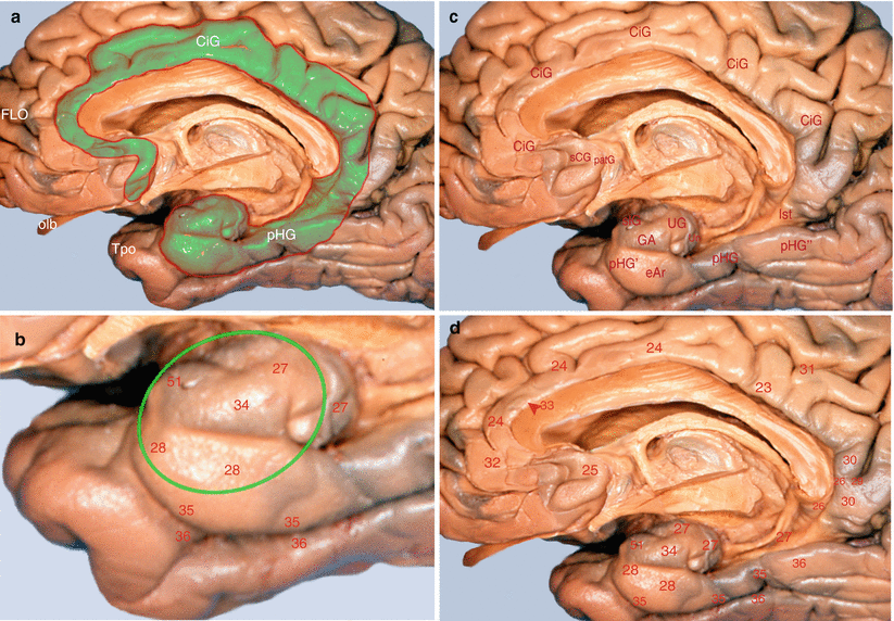

Fig. 1.1
Localisation of the amygdaloid nuclear complex in the limbic lobe. (a) Limbic lobe (green coloured) on a medial view of the right hemisphere. (b) Amygdala’s projections (green circle) on the temporal areas, according to Brodmann. (c) The gyri of the limbic lobe. (d) The Brodmann’s areas corresponding to the limbic lobe. CiG cingulate gyrus, eAr entorhinal area, GA gyrus ambiens, FLO frontal lobe, Ist Isthmus, olb olfactory bulb, patG paraterminal gyrus, pHG parahippocampal gyrus, pHG’ anterior part of pHG (piriform lobe), pHG” posterior, part of pHG, sCG subcallosal gyrus, slG semilunar gyrus, Tpo temporal pole, UG uncinate gyrus, Un uncus
1.3 Amygdala and Limbic Lobe
When Broca described the large limbic lobe, he only included, in its composition, the annular structure which surrounds the median inter-hemispheric commissures and the intermediate or diencephalic brain. This structure is essentially formed by the cingulate gyrus (referred to in the past as the pericallosal gyrus) and the parahippocampal gyrus (referred to in the past as the circonvolution of the hippocampus or fifth temporal gyrus).
Stay updated, free articles. Join our Telegram channel

Full access? Get Clinical Tree


