Fig. 9.1
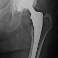
Left posttraumatic ankle arthritis. a Cobey/Saltzman view. b Anteroposterior radiograph of the left leg showing a varus deformity, with center of rotation of angulation (CORA) located at the distal third of the tibia. With permission from: Bonasia et al. [3]
In the coronal plane views, different measurements can be obtained, including the lateral distal tibial angle (LDTA), the tibial–talar angle, and the calcaneal tibial alignment. The LDTA (Fig. 9.2a) is formed by the distal tibial articular surface and the anatomical axis of the tibia and measures 89° ± 3° [3, 4]. A decreased LDTA represents a varus deformity. The tibial–talar angle (Fig. 9.2c) is defined by the tibial and talar articular surfaces of the ankle joint. When the tibial–talar angle is >10°, the joint is defined incongruent (unstable) [5]. The calcaneal–tibial alignment (measured on the Cobey/Saltzman view) is useful to confirm any varus or valgus deformities as well as to assess every talar compensation (inversion and eversion) to an abnormal LDTA. The subtalar joint can compensate 15° of eversion and 30° of inversion [4]. Evaluation of subtalar joint compensation and range of motion is important because tibial realignment (with osteotomy or TAR) may unmask a subtalar deformity. If this distal (rearfoot) deformity is not addressed, further symptoms may develop [4]. In the sagittal plane, the anterior distal tibial angle (ADTA) should be measured. The ADTA is formed by the mechanical axis of the tibia and the joint orientation line of the ankle in the sagittal plane and measures 80° ± 3° in the normal lower extremity (Fig. 9.2b) [4]. An increased ADTA represents a recurvatum deformity.

Fig. 9.2
Preoperative measurement. a The LDTA is formed by the distal tibial articular surface and the anatomical axis of the tibia (normal values 89° ± 3°). b The ADTA is formed by the mechanical axis of the tibia and the joint orientation line of the ankle in the sagittal plane (normal values 80° ± 3°). c The tibial–talar angle (α) is defined by the tibial and talar articular surfaces in the ankle joint; if it measures >10°, the joint is defined incongruent. With permission from: Bonasia et al. [3]
If an abnormal ADTA or LDTA is present (sagittal or coronal deformity), the center of rotation of angulation (CORA) is measured. The CORA is the intersection of the mid-diaphyseal line and the line starting from the middle of the joint and perpendicular to the abnormal ADTA or LDTA (Fig. 9.3) [3]. The CORA can be located at the joint line level (usually due to anatomical joint line malalignment or to ankle degeneration) or proximally (usually due to tibial deformities/fractures). Malalignment and instability should be thoroughly evaluated in the preoperative planning. Both can lead to edge-loading of the implant, polyethylene wear, progressive deformity, and high early failure rates.

Fig. 9.3
The center of rotation of angulation (CORA) is the intersection of the mid-diaphyseal line and the line starting from the middle of the joint and perpendicular to the abnormal ADTA or LDTA (LDTA in this figure). The CORA can be located proximally at the tibia (a) or at the joint line level (b). With permission from: Bonasia et al. [3]
9.3.2 Computed Tomography
Computed tomography (CT) provides accurate information about bone stock. It is not routinely performed in primary TAR, but it is often required in case of severe deformities and revision surgery and in some cases of rheumatoid arthritis.
9.3.3 Magnetic Resonance Imaging
Magnetic resonance imaging (MRI) is not routinely used preoperatively in TAR, but can be performed in case of avascular necrosis of the talus in order to confirm the diagnosis or to assess its severity and to evaluate soft tissue conditions.
9.4 Implantation Technique [3]
The patient is positioned supine, with a bump under the hip and a tourniquet on the proximal thigh. After antibiotic prophylaxis, a mid-line 10-cm skin incision centered on the joint line is performed. The superficial peroneal nerve is identified and protected throughout the whole procedure. The joint is approached through the bed of the tibialis anterior tendon or the extensor hallucis longus, and the neurovascular bundle is retracted laterally. Once in the joint, it is fundamental to achieve a correct soft tissue balancing and restore a correct tibio-talar alignment, in order to position the tibial and talar components perpendicular to the plumb line of the body. Therefore, a careful debridement of the osteophytes, synovial tissue, and redundant capsule is carried out. The medial and lateral gutters need to be debrided as well.
In a congruent varus (or valgus) ankles, the tibial cut is then performed to neutralize the LDTA and to have a reference for talar tilt reduction. In the valgus tibial deformity, more bone should be removed medially compared to laterally and vice versa in the varus tibial malalignment. The talar tilt is then reduced by subperiosteal deltoid ligament release with a Cobb elevator. If this is not sufficient, the tibialis posterior tendon is released as well.
In incongruent varus (or valgus) ankles, the LDTA is normal and no neutralizing cuts are necessary. The talar tilt is reduced with intra-articular and extra-articular soft tissue procedure, as described above.
In case of severe varus or valgus deformity (i.e., rheumatoid patients), tibial and talar bone cuts are necessary to realign the joint, with the “sculpturing technique” [6]. When performing tibial cuts with the talus still tilted, it is important to have TAR instruments that allow talar cut independent of the tibial one. Then, soft tissue procedure can be performed to achieve a rectangular space between the tibial and talar cuts.
At this point, bone cuts are completed and trial components positioned. With the trial components, ROM and stability should be checked. If limited dorsiflexion is achieved, and this is not due to component malpositioning, a percutaneous Achilles’ tendon lengthening is indicated and can be performed. If stability of the implant is not satisfactory from varus tilt, reconstruction of lateral ligaments may be necessary. Generally, if the implants are placed in anatomical alignment, with an appropriate poly spacer, instability is uncommon. If ligamentous stabilization is required, this can be performed, as for ankle instability, with anatomical repair (Broström [7] or Gould [8]), with or without auto/allograft augmentation, or with non-anatomical reconstruction tenodeses (i.e., Watson-Jones [9], Evans [10], Chrisman and Snook [11] procedures). Once a correct soft tissue balancing is obtained, definitive TAR components can be implanted.
Related posts:

Stay updated, free articles. Join our Telegram channel

Full access? Get Clinical Tree


