Fig. 8.1
Spectral Doppler echocardiographic image across the LVOT after Valsalva maneuver demonstrating a characteristic late-peaking “dagger-shaped” signal suggestive of severe dynamic LVOT obstruction, characteristic of HOCM
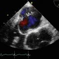
Video 8.1 Two-dimensional transthoracic echo in the parasternal view demonstrating severely hypertrophied basal interventricular septum with systolic anterior motion (SAM) of the mitral valve. The patient has a characteristic “reverse septal curvature” pattern, which is frequently seen in patients with a genetically transmitted mode of disease (AVI 5952 kb)
Video 8.2 Two-dimensional transthoracic echo with color Doppler in the parasternal view demonstrating severe turbulence of blood flow across the LVOT, due to a combination of SAM of the anterior mitral leaflet and severely hypertrophied basal interventricular septum. Notice the characteristic SAM-related posteriorly directed jet of mitral regurgitation (MR) in the left atrium (AVI 3537 kb)
Video 8.3 Four-chamber balanced steady-state free precession (b-SSFP) cine cardiac magnetic resonance (CMR) image of the same patient confirming severe basal septal hypertrophy, LVOT obstruction, and posteriorly directed jet of MR. Again, notice the characteristic reverse septal curvature pattern (AVI 1425 kb)
8.2 Case 2. Hypertensive Heart Disease of Elderly with Severe LVOT Obstruction
A 78-year-old woman presented with progressively worse exertional dyspnea. She had a long-strong history hypertension, that was not well controlled. On examination, she was found to have a harsh systolic ejection murmur at left upper sternal border that increased with Valsalva and reduced with squatting. Electrocardiogram revealed normal sinus rhythm with severe LVH. She was diagnosed with hypertensive heart disease of elderly and severe LVOT obstruction. She underwent surgical myectomy with complete relief of symptoms.

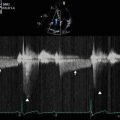
Fig. 8.2
Post-exercise spectral Doppler echocardiographic image across the LVOT in the same patient, demonstrating a characteristic late-peaking “dagger-shaped” signal, suggestive of severe dynamic LVOT obstruction
Video 8.4 Two-dimensional transthoracic echocardiogram in the parasternal view demonstrating a severely hypertrophied focal segment of the basal interventricular septum with systolic anterior motion (SAM) of the mitral valve. The patient has a characteristic “sigmoid” pattern, which is frequently seen in elderly patients with long-standing history of uncontrolled hypertension (AVI 6144 kb)
Video 8.5 Two-dimensional transthoracic echocardiogram in the apical 5-chamber view again demonstrating the characteristic sigmoid-shaped interventricular septum with systolic anterior motion (SAM) of the mitral valve (AVI 2502 kb)
8.3 Case 3. Obstructive Cardiomyopathy Without Overt Basal Septal Hypertrophy: Intrinsic Mitral Calve Pathology
A 55-year-old woman presented with positional dizziness and progressive exertional dyspnea. She had a known history of mitral valve abnormality. On examination, she was found to have a harsh systolic ejection murmur at left upper sternal border that increased with Valsalva and reduced with squatting. She also had a loud systolic precordial murmur. Electrocardiogram revealed normal sinus rhythm. She was diagnosed with obstructive cardiomyopathy without overt basal septal hypertrophy, due to intrinsic anterior mitral leaflet disease. Her anterior mitral leaflet was elongated and resulted in systolic anterior motion (SAM) and symptomatic LVOT obstruction. She underwent anterior mitral valve repair (without a myectomy) with complete relief of symptoms.

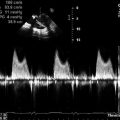
Fig. 8.3
Spectral Doppler echocardiographic image across the LVOT in the same patient, demonstrating a characteristic late-peaking “dagger-shaped” signal, suggestive of severe dynamic LVOT obstruction
Video 8.6 Two-dimensional transthoracic echocardiogram in the apical 3-chamber view demonstrating a long anterior mitral leaflet with obstructive SAM. Notice that the basal interventricular septum is normal in thickness (AVI 1709 kb)
Video 8.7 Two-dimensional transthoracic echocardiogram with color Doppler in the same apical 3-chamber view demonstrating severe turbulence of blood flow across the LVOT and characteristic severe posteriorly-directed jet of MR. However, there is no evidence of basal septal hypertrophy (AVI 0 bytes)Related posts:

Stay updated, free articles. Join our Telegram channel

Full access? Get Clinical Tree