Normal Anatomy, Including Variants, Encountered on Radiography, CT, and MR
QUESTIONS
1b How often is this type of anatomy present?
A. 10% to 20%
B. 40% to 50%
C. 80% to 90%
View Answer
1b Answer A. Codominant anatomy occurs in roughly 10% to 20% of patients.
References: O’Brien JP, Srichai MB, Hecht EM, et al. Anatomy of the heart at multidetector CT: what the radiologist needs to know. Radiographics 2007;27(6):1569-1582. Review.
Pannu HK, Flohr TG, Corl FM, et al. Current concepts in multi-detector row CT evaluation of the coronary arteries: principles, techniques, and anatomy. Radiographics 2003;23:S111-S25. Review.
2 What is the normal relationship of tricuspid and mitral valves?
A. They are located on the same level.
B. Tricuspid valve is more apically located than the mitral valve.
C. Mitral valve is more apically located than the tricuspid valve.
View Answer
2 Answer B. The tricuspid valve is more apically located than the mitral valve. This can be helpful in identifying the valves/ventricles in patients with ventricular inversion. The AV valves (tricuspid and mitral) will go with their respective morphologic ventricles (tricuspid with morphologic RV, mitral with morphologic LV).
References: O’Brien JP, Srichai MB, Hecht EM, et al. Anatomy of the heart at multidetector CT: what the radiologist needs to know. Radiographics 2007;27(6):1569-1582. Review.
Schallert EK, Danton GH, Kardon R, et al. Describing congenital heart disease by using three-part segmental notation. Radiographics 2013;33(2):E33-E46. doi: 10.1148/rg.332125086.
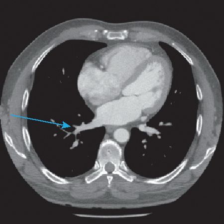
3 What is the normal relationship of the left pulmonary artery to the bronchi
A. Hyparterial
B. Eparterial
C. Isoarterial
View Answer
3 Answer A. Normal relationship of the left pulmonary artery to the left mainstem and left lobar bronchi is hyparterial (the bronchi is inferior to the bronchi). The normal relationship of the right pulmonary artery to the right main stem bronchus is eparterial (artery is superior to the bronchus). This can be used when evaluating patients with situs anomalies to determine the right and left side.
References: Lapierre C, Déry J, et al. Segmental approach to imaging of congenital heart disease. Radiographics 2010;30(2):397-411. doi: 10.1148/rg.302095112. Review.
Schallert EK, Danton GH, Kardon R, et al. Describing congenital heart disease by using three-part segmental notation. Radiographics 2013;33(2):E33-E46. doi: 10.1148/rg.332125086.
4 Which cardiac valve is the most posteriorly located?
A. Aortic
B. Mitral
C. Pulmonic
D. Tricuspid
View Answer
4 Answer B. The most posteriorly located valve is the mitral valve. The pulmonic valve is located anterior and superior to the aortic valve. The mitral valve is located posterior to the aortic valve. The tricuspid valve is the most lateral right-sided valve (typically right of the spine).
References: Lapierre C, Déry J, Guérin R, et al. Segmental approach to imaging of congenital heart disease. Radiographics 2010;30(2):397-411. doi: 10.1148/rg.302095112. Review.
Schallert EK, Danton GH, Kardon R, et al. Describing congenital heart disease by using three-part segmental notation. Radiographics 2013;33(2):E33-E46. doi: 10.1148/rg.332125086.
5 Which cardiac valve is the most superiority located?
A. Aortic
B. Mitral
C. Pulmonic
D. Tricuspid
View Answer
5 Answer C. The most superiorly located valve is the pulmonary valve. One mnemonic for remembering the pulmonary valve position is “my Pal Sal.” The pulmonic valve (PAL) is superior and anterior and to the left (SAL) relative to the aortic valve in normal anatomy.
References: Lapierre C, Déry J, Guérin R, et al. Segmental approach to imaging of congenital heart disease. Radiographics 2010;30(2):397-411. doi: 10.1148/rg.302095112. Review.
Schallert EK, Danton GH, Kardon R, et al. Describing congenital heart disease by using three-part segmental notation. Radiographics 2013;33(2):E33-E46. doi: 10.1148/rg.332125086.
6 What is the valve at the ostium of the coronary sinus?
A. Eustachian
B. Thebesian
C. Vieussens
D. Marshall
View Answer
6 Answer B. The valve at the ostium of the coronary sinus is the Thebesian valve. The eustachian valve is at the inferior vena cava. The Vieussens valve is at the junction of the coronary sinus and the great cardiac vein. The ligament of Marshall is the developmental remnant of the left superior vena cava.
Reference: Shah SS, Teague SD, Lu JC, et al. Imaging of the coronary sinus: normal anatomy and congenital abnormalities. Radiographics 2012;32(4):991-1008. doi: 10.1148/rg.324105220.
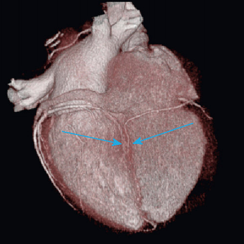
A. Crista terminalis
B. Thrombus
C. Myxoma
D. Central line
View Answer
7 Answer A. This posterior right atrial structure is the crista terminalis, which is a muscular ridge separating the muscular and smooth portion of the right atrium. It can often be mistaken for a right atrial mass/thrombus but is a normal structure. While thrombus can be associated with the crista terminalis, it would typically be larger and associated with history of central line placement. Right atrial myxoma can occur in the posterior right atrial wall but are typically larger and along the interatrial septum.
Reference: Malik SB, Kwan D, Shah AB, et al. The right atrium: gateway to the heart— anatomic and pathologic imaging findings. Radiographics 2015;35(1):14-31. doi: 10.1148/rg.351130010.
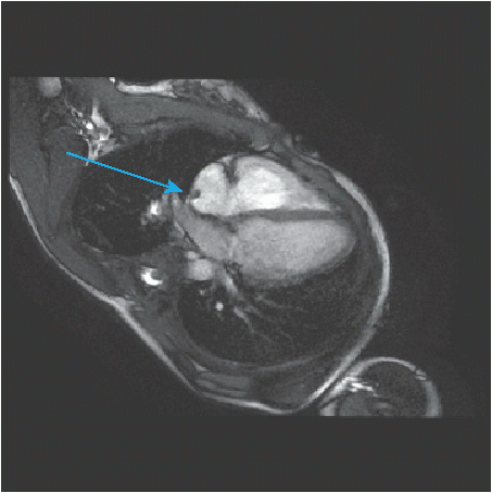
A. Right superior pulmonary vein
B. Right inferior pulmonary vein
C. Scimitar vein
View Answer
8 Answer B. The vein seen draining into the left atrium is the right inferior pulmonary vein. This can be determined due to the fact that inferior pulmonary veins drain the lower lobe, which is posteriorly located. Therefore, any vein that is approaching from the posterior lung will be draining the lower lobe and thus inferiorly located. Any vein draining anteriorly would be the superior pulmonary veins. The scimitar vein typically will drain into the right atrium/IVC.
|
Reference: Porres DV, Morenza OP, Pallisa E, et al. Learning from the pulmonary veins. Radiographics 2013;33(4):999-1022. doi: 10.1148/rg.334125043. Review.
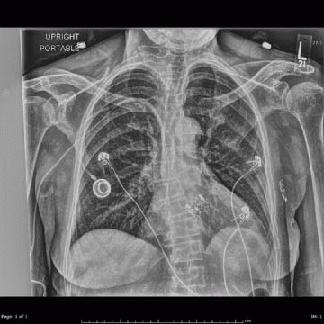
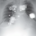
A. Aortic valve
B. Mitral valve
C. Pulmonic valve
D. Tricuspid valve
View Answer
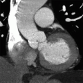
9 Answer A. The patient has undergone transaortic valve replacement (TAVR) secondary to aortic stenosis. The procedure is performed in patients who are high risk for surgery who cannot undergo an open aortic repair. The percutaneous valve is seated in the left ventricular outflow tract and ascending aorta.
References: Leipsic J, Wood D, Manders D, et al. The evolving role of MDCT in transcatheter aortic valve replacement: a radiologists’ perspective. AJR Am J Roentgenol 2009;193:3, W214-W219.
Mehlman DJ. A guide to the radiographic identification of prosthetic heart valves: an addendum. Circulation 1984;69(1):102-105.
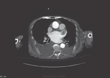
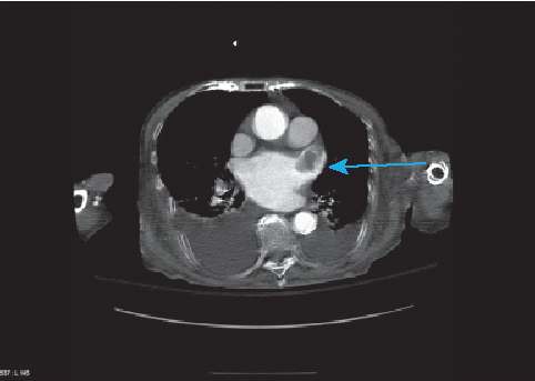
A. Aorta
B. Left atrial appendage
C. Pulmonary vein
D. Right atrium
View Answer
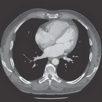
10 Answer B. The left atrium is enlarged and within the left atrial appendage is a thrombus. Thrombus can develop in the left atrial appendage in patients with recurrent atrial fibrillation and can undergo systemic embolization.
|
Reference: Garcia MJ. Detection of left atrial appendage thrombus by cardiac computed tomography. A word of caution. J Am Coll Cardiol 2009;2(1):77-79. doi: 10.1016/j.jcmg.2008.10.003.
A. Aorta
B. Brachiocephalic vein
C. Left upper lobe pulmonary vein
D. Superior vena cava
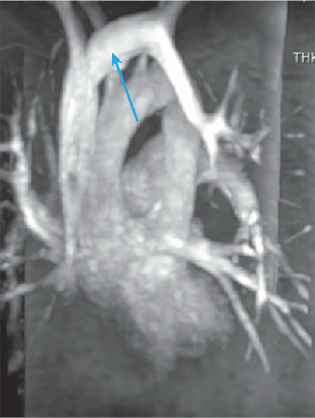
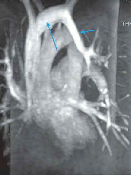
View Answer
11 Answer B. Patient has left upper lobe anomalous pulmonary venous return (arrow on the right). The anomalous vein drains into the left brachiocephalic vein and subsequently to the SVC and right atrium. The pattern of drainage creates a left-to-right shunt.
|
Reference: Dillman JR, Yarram SG, Hernandez RJ. Imaging of pulmonary venous developmental anomalies. AJR Am J Roentgenol 2009;192(5):1272-1285.
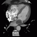
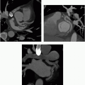
12 The image shows an oblique coronal image of the aorta. The aortic root is defined as:
A. Between the annulus and sinotubular junction
B. Between the brachiocephalic artery and aortic isthmus
C. Between the sinotubular junction and aortic isthmus
D. Between the aortic isthmus and diaphragmatic hiatus
View Answer
12 Answer A. The aortic root is defined as the segment between the aortic annulus (basal ring of the annulus and the sinotubular junction. It includes the basal ring of the annulus (aortic cusp insertion), the aortic valve cusps, and the sinuses of valsalva.
Reference: Charitos EI, Seivers HH. Anatomy of the aortic root: implications for valve-sparing surgery. Ann Cardiothorac Surg 2012;2(1):53-56.
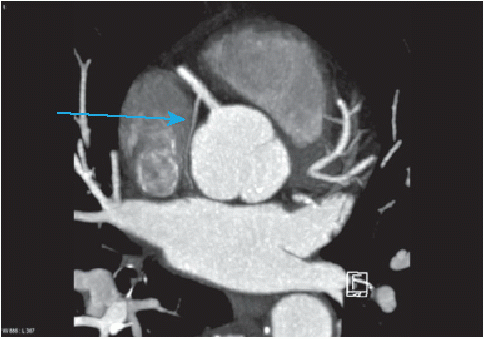
A. Anterior wall of the right ventricle
B. Infundibulum
C. Left atrium
D. Sinoatrial node
View Answer
13 Answer D. The sinoatrial nodal artery most commonly arises from the RCA and courses toward the interatrial septum. The artery can also arise from the left circumflex coronary artery.
Reference: Kini S, Bis, KG, Weaver L. Normal and variant coronary arterial and venous anatomy on high resolution CT angiography. AJR Am J Roentgenol 2007;188(6):1665-1674.
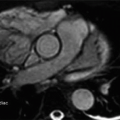
14 This short-axis balanced steady-state free precession is at the level of the midcavity of the papillary muscle. Which coronary artery typically supplies the structure the arrow is pointing to?
|
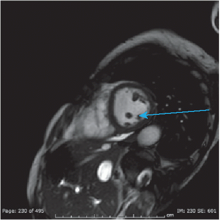
A. Left anterior descending coronary artery
B. Left circumflex coronary artery
C. Left main coronary artery
Related posts:
Stay updated, free articles. Join our Telegram channel

Full access? Get Clinical Tree


