Chapter 5 Recognizing Atelectasis
What is Atelectasis?
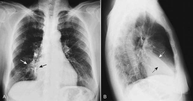
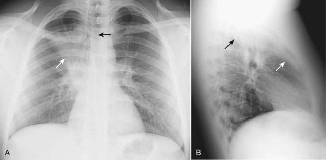
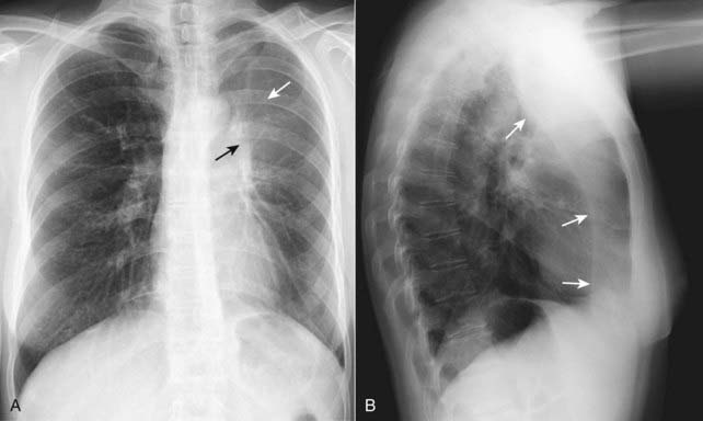
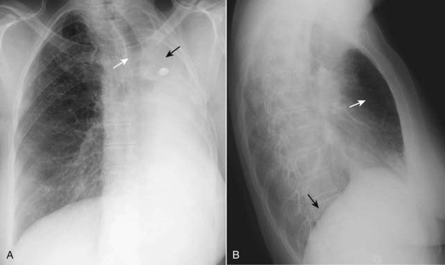
 Common to all forms of atelectasis is a loss of volume in some or all of the lung, usually leading to increased density of the lung involved.
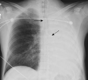
 Unless mentioned otherwise, statements in this chapter that refer to “atelectasis” are referring to obstructive atelectasis. This might be a good time to review the chart from Chapter 4 (see Table 4-1) highlighting the markedly different appearances of the thorax in a large pneumothorax and atelectasis of the entire lung (see Fig. 4-2).
Signs of Atelectasis
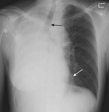
 Displacement (shift) of the mobile structures of the thorax.
• The mobile structures are those capable of movement due to changes in lung volume.
• Trachea
• Normally midline in location and centered on the spinous processes of the vertebral bodies (also midline structures) on a nonrotated, frontal chest x-ray. A slight rightward deviation of the trachea is always present at the site of the left-sided aortic knob.
• Heart
• At least 1 cm of the right heart border normally projects to the right of the spine on a nonrotated, frontal radiograph.
 Overinflation of the unaffected ipsilateral lobes or the contralateral lung.
• The greater the volume loss and the more chronic its presence, the more the lung on the side opposite the atelectasis or the unaffected lobe(s) in the ipsilateral lung will attempt to overinflate to compensate for the volume loss.
Types of Atelectasis
 Subsegmental atelectasis (also called discoid atelectasis or platelike atelectasis) (Fig. 5-7)
• Subsegmental atelectasis produces linear densities of varying thickness usually parallel to the diaphragm, most commonly at the lung bases.
• It occurs mostly in patients who are “splinting,” i.e., not taking a deep breath, such as postoperative patients or patients with pleuritic chest pain.


