Figure 9-2. Deltoid muscle tear. [A] Short-axis 12-5 MHz US image shows a discrete tear (asterisk) of the deltoid muscle (delt). [B] Corresponding long-axis 12-5 MHz US image. Hum= humerus.
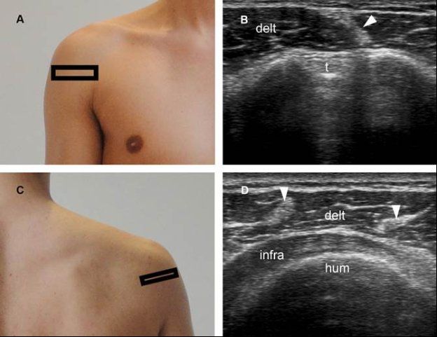
Figure 9-3. Normal deltoid muscle. [A] Positioning of the probe. [B] Corresponding 12-5 MHz US image shows normal echogenic fascia (arrowhead) mimicking fibrosis of the deltoid muscle. [C] Positioning of the probe. [D] Corresponding 12-5 MHz US image shows normal echogenic fascia (arrowheads). Delt= deltoid muscle. T= long head of the biceps brachii tendon. Infra= infraspinatus muscle. Hum= humerus.
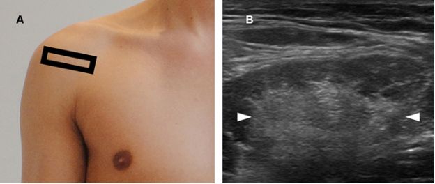
Figure 9-4. Deltoid muscle fibrosis. [A] Positioning of the probe. [B] Corresponding 12-5 MHz US image shows a hyperechoic stellate intramuscular lesion following remote shoulder contusion (arrowheads). When secondary to a strain, muscle fibrosis is usually linear; when secondary to a contusion, it may assume a stellate appearance. Stellate fibrosis must be differentiated from muscle tumors by pertinent investigation.
3.2. Fatty Atrophy
Fatty atrophy is a loosely applied term used to describe decreased volume accompanied by increased echogenicity of muscular tissue at US. Abnormal muscle echogenicity is assessed subjectively by comparison with the trapezius. With increased muscle echogenicity, the sharp contrast between the hyperechoic perimysium and hypoechoic muscle bundles becomes less distinct, leading first to the effacement and then to the disappearance of the pennate pattern (video 9-1).5 Muscle atrophy represents the end result of many causes, including aging, disuse, denervation, muscular dystrophy, and cachexia (figure 9-5). It may also occur as a result of iatrogenic injury (see figure 2-10). Detection of deltoid muscle atrophy should prompt additional evaluation of the teres minor muscle because quadrilateral space syndrome is included in the differential diagnosis (see figure 1-69).
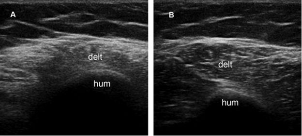
Figure 9-5. Deltoid muscle atrophy. [A] Short-axis 12-5 MHz US image shows increased echogenicity and decreased volume of the deltoid muscle (delt). [B] Comparative contralateral 12-5 MHz US image shows normal deltoid muscle in asymptomatic shoulder. Hum- humerus.
3.3. Enthesopathy
Enthesopathy is characterized by an early phase involving edema, infiltration and destructive fibrocartilage microlesions; the subsequent vascular proliferation in the subchondral bone and in the fibrocartilage causes bone erosions, reactive sclerosis and reactivation of endochondral ossification leading to enthesophytes.6
Deltoideal humeral enthesopathy is an exceedingly rare condition related to mechanical stress, typically depicted by target evaluation because of point tenderness (figure 9-6). Conversely, deltoideal acromial enthesopathy is likely a hallmark of seronegative spondylarthropathies and its detection should probably be followed by pertinent clinical and serological investigation (figure 9-7). The fact that seronegative spondylarthropathies does not affect clavicular and humeral fibrous enthesis of the deltoid muscle further support the hypothesis that the inflammatory basis of the spondylarthropathies might reflect immunity to fibrocartilage.7
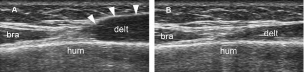
Figure 9-6. Deltoideal humeral enthesopathy. [A] Long-axis coronal 12-5 MHz US image shows thickened and hypoechoic deltoideal humeral insertion (arrowheads). [B] Comparative contralateral 12-5 MHz US image shows normal deltoideal insertion in asymptomatic shoulder. Delt= deltoid muscle. Bra= brachialis muscle. Hum= humerus.
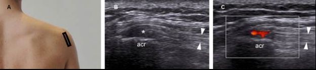
Figure 9-7. Deltoideal acromial enthesopathy. [A] Positioning of the probe. [B] Corresponding 12-5 MHz US image shows thickened and hypoechoic acromial origin of the deltoid muscle (asterisk). The tendon has normal fibrillar pattern (arrowheads). [C] Corresponding 12-5 MHz color Doppler US image shows neovascularization and indicates active disease. Acr= acromion.
Related posts:

Stay updated, free articles. Join our Telegram channel

Full access? Get Clinical Tree


