 Clinical Presentation
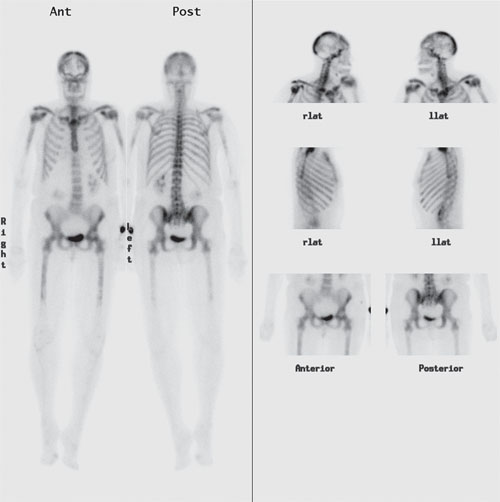
A woman with a history of breast cancer presents for re-evaluation of disease status.

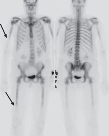
Tc99m HDP bone scintigraphy demonstrates uniformly increased radiotracer throughout the entire axial and proximal appendicular skeleton, with a sharp cutoff in uptake distal to the mid humeri and femora symmetrically (arrows). There is also an increase in the ratio of uptake in the visualized skeleton to uptake in the soft tissues, including the kidneys. Subtly decreased right breast uptake suggests a prior mastectomy. Note that the right kidney is located slightly more inferiorly in this patient than is typical.
 Differential Diagnosis
• Diffuse skeletal metastases (“metastatic superscan”): Diffusely increased osseous uptake involving the entire axial and proximal appendicular skeleton and decreased soft-tissue and renal uptake make this the correct, pathognomonic diagnosis.
• Metabolic bone disease (“metabolic superscan”): This would also have decreased soft-tissue/renal activity, but the skeletal uptake would be increased throughout the entire skeleton (head to toe).
• Multifocal Paget disease:


Stay updated, free articles. Join our Telegram channel

Full access? Get Clinical Tree


