 Clinical Presentation
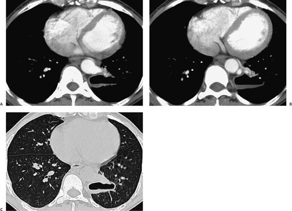
A 23-year-old man with recurrent pneumonia.
 Imaging Findings
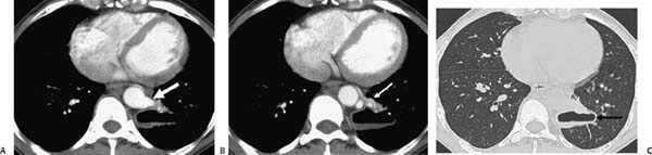
(A–C) Contrast-enhanced thoracic computed tomography. Mediastinal window (A, B) and lung window (C) images demonstrate a well-defined cavitary lesion in the left lower lobe (black arrow, C) and an abnormal vessel arising from the lateral aspect of the distal descending thoracic aorta (white arrow, A), adjacent to the cavitary lesion. An abnormal vein anterior to the aorta is also present (white arrow, B).
 Differential Diagnosis
• Pulmonary sequestration (PS):
Stay updated, free articles. Join our Telegram channel

Full access? Get Clinical Tree


