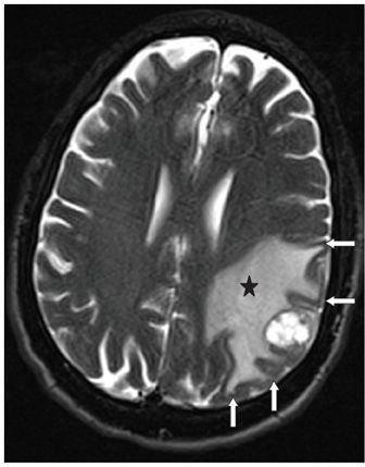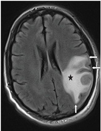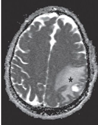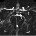


FINDINGS Figure 1-1. Axial post-contrast T1WI through the corona radiata/centrum semiovale. There is a ring-enhancing mass in the left frontoparietal junction with surrounding white matter (WM) hypointensity (star). Figures 1-2 and 1-3. Axial T2WI and FLAIR through the mass. The mass is surrounded by confluent WM hyperintensity (star) extending into the corona radiata/centrum semiovale and the subcortical WM in a finger-like fashion into the gyri sparing the cortical gray matter (GM) (arrows). The intervening sulci are effaced by mass effect. Figure 1-4. Axial ADC map through the lesion. There is confluent hyperintensity (star) of the WM around the mass (star) indicative of increased diffusivity and lack of restricted diffusion.
DIFFERENTIAL DIAGNOSIS Vasogenic edema, cytotoxic edema, encephalitis, primary malignancy, metastasis, toxoplasmosis, and abscess.
DIAGNOSIS Vasogenic edema secondary to brain metastasis.
DISCUSSION
Stay updated, free articles. Join our Telegram channel

Full access? Get Clinical Tree


