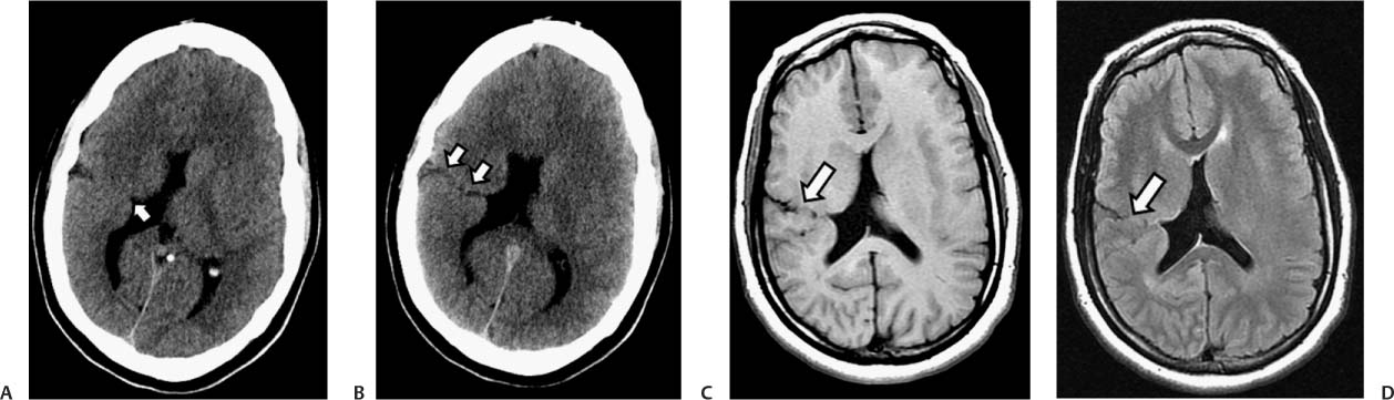
Case 10 A patient with a history of developmental delay and seizures. (A) Axial computed tomography scan of the brain demonstrates a small outpouching of the lateral wall of the right lateral ventricle, lined by gray matter (arrow). A deep sulcus is noted in the subjacent cortex. (B) A contiguous slice demonstrates a cleft with cerebrospinal fluid (CSF) attenuation through which the lateral ventricle communicates with the subarachnoid space. The defect is lined by gray matter (arrows). (C) Axial T1-weighted image of the brain show a CSF-filled cleft, lined by gray matter, between the subarachnoid space and the lateral wall of the right lateral ventricle (arrow). (D)
Clinical Presentation
Further Work-up
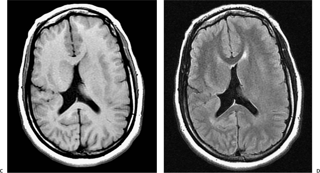
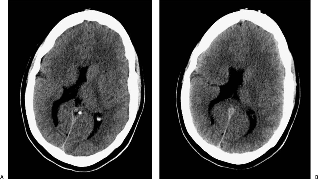
Imaging Findings
![]()
Stay updated, free articles. Join our Telegram channel

Full access? Get Clinical Tree


