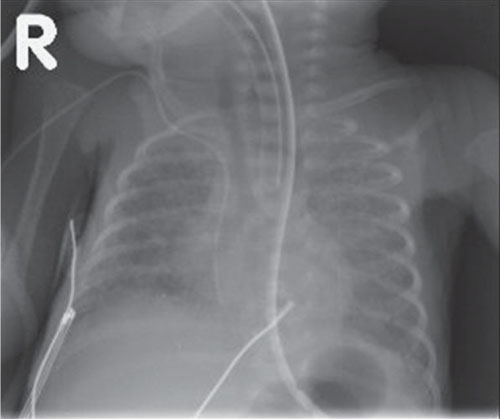
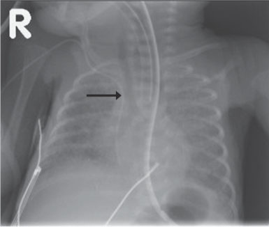
Case 10 An infant with persistent respiratory distress and poor breath sounds despite intubation. Frontal chest radiograph demonstrates underinflated lungs and the endotracheal tube paralleling the course of the esophageal tube, with the tracheal air column clearly positioned separately and to the right (arrow). • Esophageal intubation: An endotracheal tube positioned in the esophagus can be very difficult to detect on frontal radiographs, but in this case seeing the endotracheal tube positioned lateral to the trachea establishes the diagnosis.
Clinical Presentation
Imaging Findings
Differential Diagnosis
Stay updated, free articles. Join our Telegram channel

Full access? Get Clinical Tree


