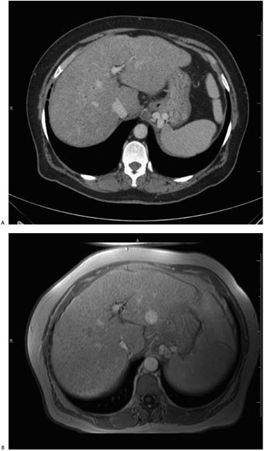
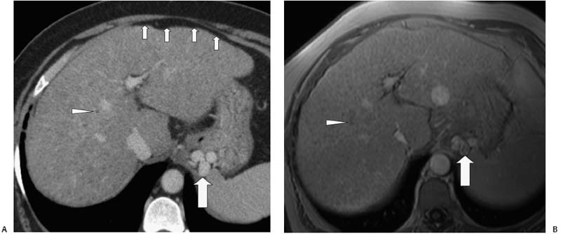
Case 10 A 63-year-old woman presents with weight loss and nausea. (A) Infused abdominal computed tomography (CT) shows hypertrophy of the left lobe of the liver; reduction of volume of the right lobe; irregular liver contour (small arrows); diffusely heterogeneous reduction in liver density (right > left); innumerable small, hypodense parenchymal nodules (arrowhead); and a cluster of enhancing structures in the gastric wall, consistent with varices (large arrow). (B) Infused T1-weighted magnetic resonance imaging (MRI) shows the liver to be abnormally hypointense relative to spleen, resulting from hypointense nodules (arrowhead). Mixed signal in gastric varices results from slow, turbulent flow (arrow). • Cirrhosis:
 Clinical Presentation
 Imaging Findings
 Differential Diagnosis
![]()
Stay updated, free articles. Join our Telegram channel

Full access? Get Clinical Tree


