CASE 10
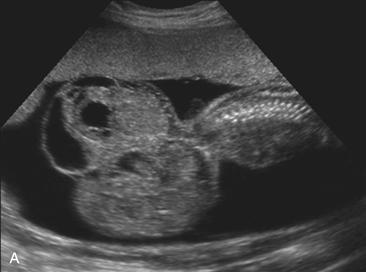
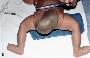
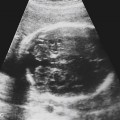
Used with permission from McGahan JP, et al: Fetal abdomen and pelvis. In McGahan JP, Goldberg BB [eds]: Diagnostic Ultrasound, 2nd ed. New York: Informa Healthcare USA, 2008; 1316. Courtesy of Marshal Swartz, MD.
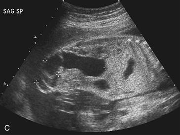
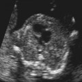
Used with permission from McGahan JP, et al: Fetal abdomen and pelvis. In McGahan JP, Goldberg BB [eds]: Diagnostic Ultrasound, 2nd ed. New York: Informa Healthcare USA, 2008; 1316.
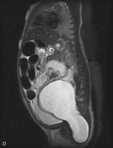
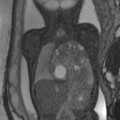
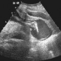
History: A patient from an outside institution presents with an ultrasound scan showing a fetal mass and undergoes a second ultrasound examination.
1. What should be included in the differential diagnosis of Figure A? (Choose all that apply.)
C. Omphalocele
2. Which of the following statements concerning sacrococcygeal teratoma is not true?
A. Incidence is approximately 1:40,000.
B. Most prenatally detected sacrococcygeal teratomas are malignant.
C. Sacrococcygeal teratomas are associated with a female-to-male ratio of approximately 4:1.
D. Few sacrococcygeal teratomas are entirely internal within the sacrum.
3. Which of the following statements concerning the ultrasound appearance of sacrococcygeal teratoma is not true?
A. Fetal MRI may be helpful to detect the internal presacral components of sacrococcygeal teratoma.
B. Sacrococcygeal teratomas are often associated with an abnormal karyotype.
C. Color Doppler ultrasound may show a highly vascular mass with large solid teratomas.
D. These tumors may be cystic, solid, or mixed.
4. Which of the following is not
Stay updated, free articles. Join our Telegram channel

Full access? Get Clinical Tree


