Case 10
 Clinical Presentation
A 32-year-old man with hard testicular swelling on the right.
 Imaging Findings
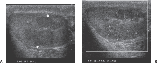
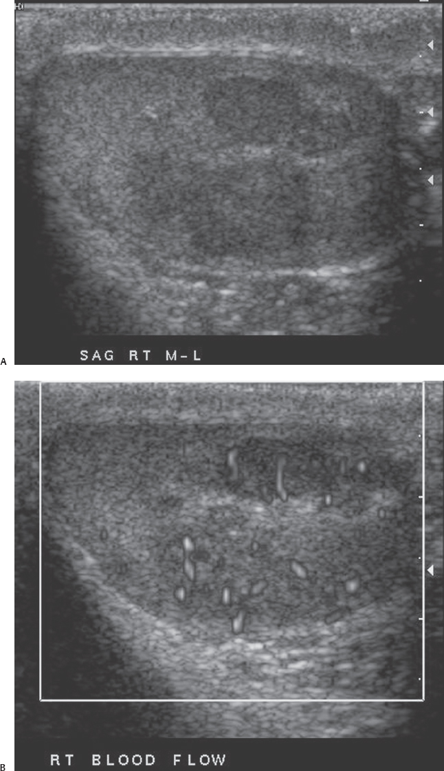
(A) Longitudinal gray scale sonographic image of the right testicle shows a well-defined, lobular, hypoechoic homogeneous mass (arrows). No calcifications or cystic areas are seen. (B) Power-flow image at the same level as Figure A shows increased blood flow within the substance of the mass (arrowhead).
 Differential Diagnosis
• Testicular neoplasm: All solid-appearing masses of the testicle should be considered neoplastic unless there is overwhelming evidence suggesting the opposite. Of the testicular neoplasms, seminoma is usually well defined, hypoechoic, and homogeneous. Internal vascularity is seen in tumors > 1.5 cm in diameter. However, smaller tumors are most often hypovascular.
• Testicular hematoma:
Stay updated, free articles. Join our Telegram channel

Full access? Get Clinical Tree


