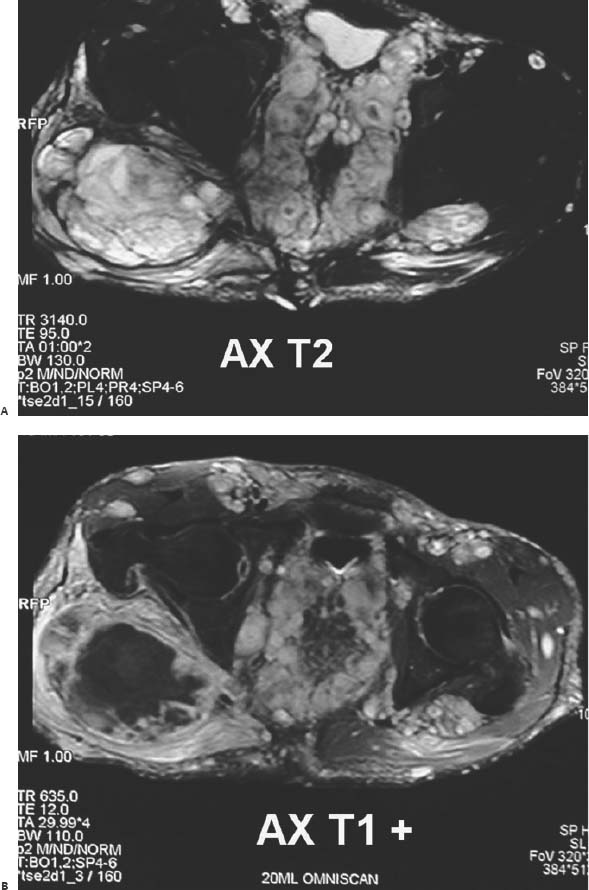
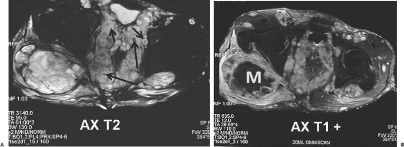
Case 100 The patient is a disfigured 32-year-old man with an enlarging gluteal mass on the right side. (A,B) Axial magnetic resonance (MR) images of the pelvis reveal multiple rounded masses along the expected course of the lumbosacral plexus with peripheral high T2-weighted MR signal intensity (short arrows) and central low T2 signal intensity (long arrows) consistent with the “target sign.” A dominant mass (M) is seen over the right gluteal region with a central area of necrosis containing high T2 signal intensity without contrast enhancement.

 Clinical Presentation
 Imaging Findings

Stay updated, free articles. Join our Telegram channel

Full access? Get Clinical Tree


