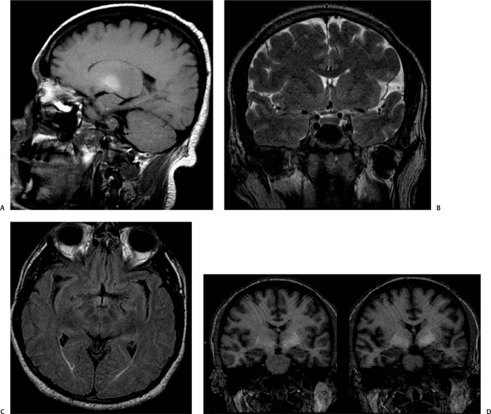
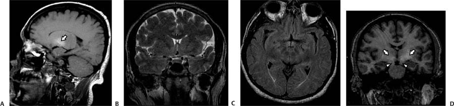
Case 100 A 37-year-old man with cirrhosis presents with rigidity and an acute confusional state. (A) Sagittal T1-weighted image (WI) shows increased T1 signal in the globus pallidus (arrow). (B) Coronal T2WI shows normal signal in the globus pallidus. (C) Axial fluid-attenuated inversion recovery (FLAIR) image shows no abnormal signal. (D) Coronal T1WI shows increased signal bilaterally in the globus pallidus (arrows) and in the substantia nigra (arrowheads). • Hepatic encephalopathy:
Clinical Presentation
Imaging Findings
Differential Diagnosis
![]()
Stay updated, free articles. Join our Telegram channel

Full access? Get Clinical Tree


